Generality
The humeral trochite is the lateral bone process of the proximal end of the humerus. Said in simpler terms, it is the characteristic protuberance located on the outer side of the upper portion of the arm bone.

The humeral trochite is an anatomically important bone region, since on it find three of the four muscles that make up the known rotator cuff of the shoulder.
The rotator cuff of the shoulder is a muscle-tendon complex essential for the main movements of the arm.
Like most of the bony elements of the human body, the humeral trochite can be subject to fractures.
What is Homeric Trochite?
The humeral trochite is an important protuberance (or process) of the humerus, that is the bone of the arm .
In human anatomy, the humeral trochite is also known as the large tubercle of the humerus .
Humerus revision
In the human being, the humerus is the even bone that constitutes the skeleton of the arm; the arm is the anatomical portion of each upper limb, which runs from the shoulder to the elbow.
The humerus belongs to the category of long bones and takes part in the formation of two important joints: the glenohumeral articulation of the shoulder, above, and the articulation of the elbow, below.
Like any long bone, the humerus can be divided into three main portions, known as: proximal end (or proximal epiphysis ), body (or diaphysis ) and distal end (or distal epiphysis ).
The proximal end of the humerus is its upper portion, ie that which borders the shoulder and which takes part in the glenohumeral joint; the body of the humerus is its central portion, ie that interposed between the proximal end and the distal end; finally, the distal end of the humerus is its lower portion, ie the one that borders the forearm and that forms the elbow joint.
Functionally, the humerus is important because:
- It takes part in joints fundamental for the movements of the entire upper limb, in particular arm;
- It receives the muscles that support the movements of the aforementioned joints;
- In young children, it is a support for four-legged locomotion.
Anatomy
Introduction: to fully understand the following anatomical description of the humeral trochitis, readers are advised to look at the figures relating to the humerus.
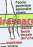
The humeral trochite is the bone process located on the proximal end of the humerus, in a lateral position with respect to the so-called humeral head, and in a posterolateral position with respect to the so-called minor tubercle .

What are the head of the humerus and the minor tubercle?
- Head of the humerus: observing the humerus from above, is the very first portion of this bone.
Projected in the medial direction, the head of the humerus is a semi-spherical protuberance, which has a smooth surface with a cartilaginous nature.
Its function is to articulate the humerus at the glenoid cavity (or glenoid fossa) of the scapula, forming the glenohumeral joint of the shoulder.
- Tubercle minor: it is a bone process (or bony protuberance) of reduced dimensions, in medial and anterior position with respect to the humeral trochite.
Equipped with a single face, the anterior one, the minor tubercle has the important task of inserting the terminal head of the rotator cuff muscle known as the subscapularis muscle .
The upper and lateral surfaces
On the humeral trochite two surfaces are recognizable, to which the anatomists have assigned the names of upper surface and lateral surface, clearly referring to the position of one with respect to the other.
Starting from the description of the less important, ie the lateral surface, this is convex and rough to the touch, and is in continuity with the lateral surface of the body of the humerus.
The upper surface, on the other hand, appears as a region only slightly rounded, on which three flat areas are distinguished, called upper facet, median facet and lower facet .
Named according to their position, the three facets of the upper surface of the humeral trochite cover a very important function, which will be discussed in the following chapter.
Intertubercular groove
Anteriorly (therefore on the front part of the humerus), to delimit the extension of the humeral trochite there is an evident fissure, which the anatomists call intertubercular sulcus .
The intertubercular sulcus marks, in fact, the border between the humeral trochite and the adjacent minor tubercle; as well as a borderline, it is also important for two other reasons:
- Inside it runs the tendon belonging to the long head of the so-called biceps brachial muscle ;
- On its superficial margins (the so-called lips of the intertubercular sulcus), tendons of the pectoralis major, large round and large dorsalis are inserted.
Function
The humeral trochite is an anatomically important bone region, because the tendon of the supraspinatus muscle, the tendinus of the sub-spinal muscle and the tendon of the small round muscle are located on the upper, middle and lower facets, respectively.
As perhaps some readers will already know, these three muscles just mentioned belong to the so-called rotator cuff of the shoulder .
Upper face → Supraspinatus muscle
Middle face → Muscle in the back of the mouth
Lower face → Small round muscle
Rotator cuff: what is it and what functions does it cover?
The rotator cuff is that important muscle-tendon complex of the shoulder, which gives stability to the latter and allows movements of extension, internal rotation, external rotation, abduction, adduction and circling of the arm .
To compose the rotator cuff are, to be precise, four muscles and their relative tendons.
Already mentioned all of them in the course of this article, the muscles in question - it is worth remembering given the important functions they perform - are:
- The supraspinatus muscle (or supraspinatus or supraspinatus), in an upper position.
- The subscapularis muscle, in anterior position (NB: for its insertion, see the previous study on the minor humerus tubercle).
- The spinal muscle (or infraspinatus) and the small round muscle, in the posterior position.
| Rotator cuff muscle | Function |
| Supraspinatus muscle | circling |
| Subscapularis muscle | Internal rotation of the arm circling |
| In spindle muscle | External rotation of the arm |
| Small round muscle | Arm extension Internal rotation of the arm External rotation of the arm circling |
diseases
Because of the position it occupies, the humeral trochite can be subjected to fractures both of compound type (therefore, of contained gravity), and of decomposed type (therefore significantly relevant).
In most cases, fractures of the humeral trochitis are the result of accidental falls, in which the victim has impacted the ground directly with the lateral portion of the shoulder or with the hypertensive arm forward.
On the occasion of more serious fractures of the humeral trochitis, lesions of one or more elements of the rotator cuff and / or the so-called dislocated shoulder may be added to bone fragmentation.
Compound fracture of the humeral trochite
The compound fracture of the humeral trochite is an injury of contained gravity, in which the broken bone portion retains its natural anatomical position.
Responsible for shoulder pain and limiting arm movements, episodes of compound fracture of the humeral trochite are difficult to diagnose by standard X-ray examination; in fact, to identify them, a more exhaustive diagnostic imaging test is needed, such as magnetic resonance imaging.
Very common in those who are victims of bad falls from bicycles or skis, the compound fracture of the humeral trochite does not usually require the use of surgery, but only requires:
- Immobilization, by plastering, of the shoulder-arm complex of the affected upper limb;
- Rest of the shoulder-arm complex;
- Taking painkillers, especially in the early stages of therapy;
- Physiotherapy once the fracture has been consolidated.
Although they are not serious injuries, compound fractures of the humeral trochite can take some time to heal; moreover, those who are victims of it can continue to feel a slight pain or discomfort for several months after consolidation.
Displaced fracture of the humeral trochite
The displaced fracture of the humeral trochite is an injury of significant severity, in which the bone portion subject to breakage has undergone a displacement with respect to its natural anatomical position.
Easily detectable by X-rays, episodes of displaced fracture of the humeral trochite cause intense pain, severely limit the ability to move the arm and are often associated with dislocations of the shoulder and / or lesions of the rotator cuff (with logical repercussions on the symptomatology ).
Displaced fractures of the humeral trochitis only heal with surgery. The surgery, in fact, is the only treatment that allows bone consolidation and allows the repair of a possible lesion of the rotator cuff or the restoration of any shoulder dislocation.
Clearly, after the surgery, the patient will undergo immobilization of the affected upper limb, he will have to rest and, once the fracture has been consolidated, he will have to follow a series of physiotherapy treatments.
Recovery times from a displaced fracture of the humeral trochite are very long and the patient may be affected by the injury for several years to come.