Generality
The navel is a cupoliform dimple located on the anterior surface of the abdomen, along the median line.

Immediately after the birth, when the child is now ready for an independent life, the umbilical cord (now useless) is severed; the residual stump undergoes necrosis and detaches spontaneously, generally at the end of the second week of life.
The navel therefore represents a scar formation and, after a gradual retraction process, assumes the appearance of a depression, circumscribed by a cutaneous ring ( umbilical rim ), at the end of which protrudes (a node or umbilical nipple ).
For anatomical constitution, the navel represents a point of least resistance of the abdominal wall. This is evident in pregnancy, ascites and umbilical hernia, conditions in which the cavity can be reduced, while the rim expands.
The navel may be the site of numerous pathological processes : among these are the infalite (phlogosis of the umbilical region), the hernia and the fistulas.
The navel as a weapon of seduction
In many cultures, the navel is considered a real weapon of seduction: just think, for example, of belly dancing or the conformation of the sari, the traditional dress of Indian women, which leaves this part of the body uncovered.
This erotic attribution dates back to ancient times: in Greek mythology, Onfale (feminine of "onfalòs", which in ancient Greek means "navel") was the queen of seduction.
The reason why the navel represents an erogenous zone is the subject of debate among scholars: some argue that its form unconsciously evokes other bodily orifices; others believe that exhibiting and discovering this part is a sign of fertility and predisposition to pregnancy.
Features
seat
The navel is found on the alba line (a thin fibrous membrane between the margins of the two rectus muscles, formed by the aponeurosis of the external oblique muscle, the internal oblique muscle and the transversus muscle), located at the level of the median portion of the abdomen .

Appearance
The navel looks like a cupoliform dimple surrounded by a cutaneous ring ( umbilical rim ), in the bottom of which a relief (the so-called " node ") protrudes, more or less evident in the central part. At the apex, this prominence presents the umbilical scar, separated from the rim by a circular groove. The peritoneum is found in this area within the body.
The navel can be hollow (more common form, similar to a depression) or pronounced (rather rare, the groove tends to escape from its cavity). The bottom can be, instead, smooth or crossed by small furrows.
Usually, the color of the navel has the same complexion as the rest of the body. In some subjects, however, the pigmentation of this dimple can vary from rosy to red, from brown to dark brown.
Line nigra in pregnancy
During gestation, a dark vertical sign may appear that extends from below the breast to the pubic area, crossing the navel at the median portion of the abdomen. This phenomenon is common and depends on the action of estrogen hormones, which stimulate the production of melanin, causing hyperpigmentation of the linea alba.
The so-called "nigra line" appears, generally, starting from the second trimester of a pregnancy on the abdomen and usually disappears within a few weeks of the birth, in a completely spontaneous way.

Linea Nigra - Image from Wikipedia.org
How the navel is formed
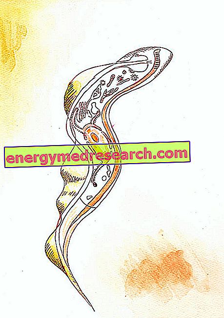
The navel corresponds to the insertion point of the umbilical cord (or funiculus).
This channel joins the fetus to the placenta and, during the intrauterine life, allows the complete metabolic support from the maternal organism.
During development, in fact, the child is completely dependent on the mother's organism for nourishment, respiration and elimination of waste. The umbilical cord contains the allantois, the blood vessels (two arteries and an umbilical vein) and the vitelline duct.
From the moment of stabilization of the embryonic appendages until birth, the fetus remains firmly joined to the uterine wall through the funicular structure, remaining suspended in the liquid contained in the amniotic sac.
The navel therefore originates in the form of an umbilical peduncle following the implantation of the embryo on the uterine wall and the development of extra-embryonic membranes.
After birth, the umbilical cord that unites the child to the mother is severed by the doctor and the remaining end ( stump ) is tied into a small knot. Within a short time, the stump begins to heal and dries, detaching itself definitively from the child's belly, leaving no residue. Thus the navel is formed.

Ligation of the umbilical cord 9 days after delivery
The characteristic appearance of this dimple does not depend, therefore, on genetic factors, but simply on the subsequent tissue healing process .
The navel varies from person to person: it can be more hollow or pronounced depending on the way in which the wound is healed, the ability of the doctor who knotted the umbilical cord after childbirth and how much residual limb was left (if too much, it will stick outwards). For this reason, there are various sizes, shapes and colors.
Treatment of the umbilical stump
The navel is formed by a gradual healing process. After childbirth, the umbilical cord is severed and the residual stump is knotted and bandaged with sterile gauze. When the latter remains completely dry, it will come off the abdomen of the newborn spontaneously (generally, within two weeks of birth), leaving a protuberance, destined, then, to flatten out completely.
In this period, in order to better manage the natural fall of the umbilical cord residue, its daily hygiene must be scrupulously treated: until it is completely healed; in fact, the abutment remains a potential gateway to the organism for various external agents.
During the period necessary for the spontaneous fall, it is therefore necessary to keep the umbilical stump clean, cleaning it with water and mild soap, if dirty or sticky, using a cotton swab or gauze. After this operation, the area must be dried carefully by placing an absorbent cloth on it or by aerating it with a piece of paper, used as if it were a fan. When changing the diaper, the umbilical stump should be left outside the area (so that it is exposed to the air and heals faster), folding the absorbent slightly downwards.
In its drying process, the umbilical stump will take on various colors: from green-yellowish to brown-black. Even if this residue seems to be attached only by a dry cord, it should never be pulled, but it is necessary to wait for it to fall by itself.
Redness in the umbilical area (with or without edema), continuous blood loss or yellowish secretion (pus) may indicate an alteration in the healing process. In this case, it is important to notify the pediatrician, as there may be an infection that should be treated promptly.
omphalitis
The navel and the surrounding tissues can be the site of inflammatory processes, called omphalitis.
These inflammations are frequent especially in newborns, due to the infection of the wound that remains after the fall of the umbilical stump; this de-epithelialized area is in fact susceptible to the potential attack of pathogenic microorganisms, such as streptococci and staphylococci. In adults, defalite can be caused, instead, by bad hygiene or by the particular anatomical conformation of the navel, which makes it difficult to clean.
The inflammation is manifested by redness, swelling, burning, tenderness and pain localized in the umbilical region. These symptoms are often accompanied by foul-smelling, purulent and continuous secretions, which make the navel always moist.
If treated properly, the condition disappears very quickly. In rare cases, however, the disorder can evolve severely, leading to the formation of cysts that require surgical removal or even giving rise to septicemia.
The management of malaria involves the topical application of local antiseptics, medicated gauzes and disinfectant ointments to eliminate the infectious process; if particularly serious, the doctor may prescribe a systemic antibiotic therapy.
Umbilical hernia
A fairly frequent alteration of the navel is the hernia. This condition can be established following the bending of an intestinal tract through the umbilical scar (weak point of the abdominal wall).

In adulthood, umbilical hernias can be observed as a result of obesity, multiple pregnancies, excessive physical exertion or lifting of heavy loads.
This anomaly can be easily reduced by surgery, repositioning the intestinal loop within the abdomen. If not treated properly, the hernia can become strangled or imprisoned.
Belly button extroflexion
Due to its anatomical constitution, the navel represents a point of lesser resistance of the abdominal wall : therefore, in some morbid conditions (as in ascites), the dimple suddenly disappears and the umbilical groove may even evert.
Even in pregnant women, the navel cavity tends to subversion from its cavity due to the pressure of the fetus on the belly, but it normally re-enters after giving birth.
Other navel pathologies
- In the adult, the navel may be involved in candida intertriginium, psoriasis and scabies .
- Sebaceous cysts are also frequent, which often undergo inflammation. The navel can also be the site of keloids of the umbilical scar, dermoid cysts, polyps, seborrheic keratoses, dermatofibromas, eczema or other dermatoses affecting the skin folds (eg mycosis, etc.).
- The umbilical fistula is a pathological process that recognizes various origins. This complication may be congenital or may result from intestinal diseases (such as helminthiasis), gallstones, and tuberculous peritonitis.
- Another disorder is umbilical endometriosis, a rare disease characterized by the presence of ectopic endometrial tissue (ie in an anomalous location), which assumes the proliferative and functional attitudes that occur in the eutopic one of the uterine wall. In this case, in women of child-bearing age, it is possible to develop erythema in the umbilicus and neighboring areas, sometimes with blood loss from these sites, at the same time as the appearance of menstrual flow.
- The navel can also be a localization of neoplastic processes : a sign indicative of this occurrence is the " nodule of Sister Mary Joseph", a subcutaneous lesion of solid consistency that can arise in the presence of metastases deriving above all from intestinal malignant tumors (such as gastric adenocarcinoma). Generally, this nodular formation does not cause any pain, but it can give rise to pus and can have a blue-violet, brown-reddish or whitish color.