The cytoplasmic fluid inside the muscle cells is largely occupied by the myofibrils, which constitute the contractile component.
Each muscle fiber is made up of about 1000 myofibrils, wrapped by the sarcoplasmic reticulum; the myofibrils extend over the entire length of the fiber and are organized in long longitudinal bundles.
Each myofibril has a thickness between 0.5 and 2 µm, for a length ranging from 10 to 100 microns (1 micron = 1/1000 of mm.).
As anticipated, the myofibrils are surrounded by the sarcoplasmic reticulum, a complex system of vesicles and tubules that gives rise to the sarcotubular system. The purpose of this structure is to accumulate the calcium necessary for contraction.
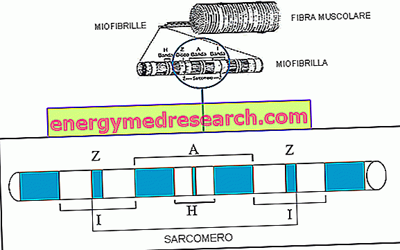
Entering more and more into the microscopic, we discover that myofibrils are in turn composed of parallel myofilaments, which are of two types: thick and thin. It is also possible to observe a characteristic streak along the major axis of the myofibril, due to the regular alternation of light and dark bands.
- The dark bands are called bands or disks A
- The light bands are called bands I
- Each band I is divided into two by a line Z
- Each band A is divided in two by a stria, called H, placed in its central part.
The myofibril tract between two adjacent Z lines
(1/2 band I + band A + 1/2 band I)
takes the name of SARCOMERO
The sarcomere is the structural and functional unit of the myofibril, that is to say the smallest unit of muscle capable of contracting.
Within the single myofibril the various sarcomeres follow one after the other, as if to form a high pile of cylinders. Furthermore, in the muscle, the fibers are arranged parallel, so that the respective sarcomeres are aligned. In other words, next to a line Z of a myofibril there is always a line Z of the adjacent myofibril; this symmetry means that as a whole, all the muscle fiber appears cross-striped.
Myofilaments
Observed under the electron microscope, each sarcomere appears to be formed by a bundle of filaments, arranged longitudinally and parallel to each other. The components of these myofilaments are two proteins, called actin and myosin.
At the center of each sarcomere is about a thousand thick filaments, consisting of myosin. At their ends, these protein molecules draw relationships with thin filaments, made up of another protein, actin.
In a skeletal type muscle cell these contractile elements (thick and thin filaments) are placed in register and are partially interdigitated (superimposed).
- The bundle of thick filaments (myosinic) is located at the center of the sarcomere and constitutes the band A;
- The bundle of thin filaments, constituted by actin, is located at the poles of the sarcomere and constitutes the two half bands I, which reach up to the Z disks.

This complex structure is the basis of muscle contraction, made possible by the sliding of thin filaments over thick ones.
During the contraction, the sarcomere is shortened by approaching the two Z-strands:
while the length of the filaments and of the A band remains unchanged there is a reduction of the I band and the H band.
The generalization of the phenomenon determines the shortening of myofibrils, muscle fibers, fascicles and the entire muscle. It is interesting to note that each sarcomere can shorten up to 50% of its length at rest.
During muscle contraction the actomyosin bridges are continuously formed and dissolved, provided that a sufficient amount of calcium ions and ATP is available; we will address this issue better in the next article.

THE VOLTAGE DEVELOPED BY A MUSCULAR FIBER IS DIRECTLY PROPORTIONAL TO THE NUMBER OF TRANSVERSAL BRIDGES FORMED BETWEEN THICK AND THIN FILAMENTS.
As a result, a muscle that is too long or too tight develops less force than a muscle that contracts starting from an optimal degree of elongation.

- A) there is no active force since there is no contact between the myosin heads and actin
- Between A) and B): there is a linear increase in the active force due to the increase of actin binding sites for myosin heads
- Between B) and C): the active force reaches its maximum peak and remains relatively stable; in this phase, in fact, all myosin heads are related to actin
- Between C) and D): the active force begins to decrease as the overlapping of the actin chains reduces the binding sites available for the myosin heads
- E): once myosin collides with disk Z there is no active force since all myosin heads are attached to actin; furthermore, myosin is compressed on the Z disks and acts as a spring, opposing the contraction with a force proportional to the degree of compression (hence muscle shortening)