The trochanter is a bony protrusion of the femur.

The two trochanters of the femur. The small trochanter, circled in red, is located in the inner region; the large trochanter, circled in orange, is located in the outer region of the femur.
The femur is the longest and largest bone in the human body, which forms the skeleton of the thigh.
Specifically, for each femur, two trochanters can be recognized: a large trochanter (more voluminous) located externally and a small trochanter (smaller) situated internally.
Trochanters act as an insertion point for several muscles involved in hip and thigh movement.
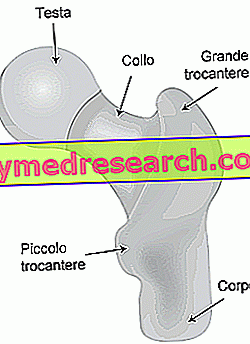
The femur and trochanters
The femur is the longest, largest and most resistant bone in the human body. It belongs to the category of so-called long bones and as such, from the anatomical point of view, presents:
- an elongated central part, called body or diaphysis :
- two ends, called epiphyses :
- the upper extremity (proximal epiphysis) presents:
- a head : it has the shape of an hemisphere (2/3 of sphere), whose rounded part (covered with cartilage) articulates with the acetabulum of the iliac bone to form the hip joint (or coxofermoral articulation) ; the cartilaginous lining is missing in a dimple (fovea capitis) where it inserts the round ligament of the femur, which serves to hold the coxofemoral joint in position
- a neck : it is the portion of connection between the head and the proximal diaphysis; Cylindrical in shape, it is about 5 centimeters long and forms with the diaphysis an angle that varies from 120 ° to 145 ° (it is generally lower in the female than in the male, having this a wider basin). The neck has a large number of channels for the passage of blood vessels.
- the lower extremity (distal epiphysis) has two large condyles and articulates with the tibia to form the femoral-tibial articulation and with the patella to form the patellofemoral joint; both are part of the knee joint .
- the upper extremity (proximal epiphysis) presents:

In the upper part of the diaphysis, at the base of the neck, two rather pronounced bony reliefs, called trochanters, can be recognized.
- The large trochanter is located laterally to the neck
- The small trochanter is located internally, behind and below the neck.
Some sources consider the trochanter regions of the proximal epiphysis.
Great trochanter
The great trochanter is a large bony prominence, quadrangular in shape, palpable on the lateral surface of the hip.
It is located above the body of the femur and marks its upper limit. It develops in the area where the body joins the neck of the femur, in a lateral position.
Later, the great trochanter is rounded and delimits a deep depression on the inner surface, called the trochanteric fossa. On the lateral wall of this fossa there is an evident oval dimple for the insertion of the external obturator muscle.
On the anterolateral surface, the large trochanter has an elongated crest due to the insertion of the small gluteal muscle. Another similar crest is found on the lateral surface, but in a more posterior position; it acts as an insertion of the gluteus medius. Between these two points the great trochanter is palpable.
On the inner-upper part of the great trochanter, just above the trochanteric fossa, there is a small area for the insertion of the internal obturator and the twin muscles; immediately above and behind it the edge of the trochanter inserts into the piriformis muscle.

Figure: Posterior view of the upper extremity of the femur.
Small trochanter
The small trochanter is smaller than the large trochanter. Its shape is conical and squat, blunt. It protrudes in the opposite position from the large trochanter, then in the inner region of the femur, just below the junction with the neck.
Under the small trochanter there is the surgical neck of the femur which - according to some sources - marks the end between epiphyses and diaphysis.
The small trochanter constitutes the insertion site for the combined tendons of the large psoas and iliac muscles (called ileo-psoas).
Between the two trochanters extend:
- anteriorly the intertrochanteric line
- posteriorly the intertrochanteric crest
These bony ridges separate the body from the neck of the femur and represent lines of connection between the two trochanters.
Intertrochanteric line
This bone crest is located on the anterior surface of the upper margin of the body. It originates from a tubercle placed on the front surface of the base of the great trochanter and descends to a position just anterior to the base of the small trochanter. At the bottom, continue with the pectineal (or hopeful line) that curves medially under the small trochanter and around the body of the femur until it joins with the medial lip of the asphalt line in the posterior part of the femur.
The intertrochanteric line provides insertion to the joint capsule on the front face of the bone.
Intertrochanteric crest
This bone crest is located on the posterior surface of the femur and descends from the posterior margin of the large trochanter to the base of the small trochanter. It appears as a smooth bone crest with a prominent tubercle (called square tubercle) located in the upper half, which provides the insertion for the square muscle of the femur.
Fractures of the trochanters
Femur fractures involve - in most cases and especially in the elderly - the neck of the femur. Indeed, after the age of 70, fractures of the neck of the femur are the most frequent fractures, both in men and above all in women (for whom the risk is greater).
This is because on one side the proximal end of the femur is often undermined by osteoporosis and on the other because in the elderly the fall modalities tend to expose this area to trauma.
The most serious consequence of these fractures of the femoral neck is the possible blood interruption of the femoral head. In fact, spraying of the head and neck mainly depends on a ring of arteries located at the base of the neck.
In the absence of blood circulation, the femoral head undergoes necrosis, ie it "crumbles" little by little. In the elderly, a fracture of the neck of the femur almost always leads to the implantation of a total hip prosthesis, while in younger patients an attempt is made to preserve the joint by restoring the fracture by osteosynthesis.
Fractures of the proximal femur have been divided into various categories based on the area where they occur. In detail they are presented:
- intertrochanteric fractures;
- fractures of the neck of the femur;
- subtrochanteric fractures;
- fractures of the great trochanter.
Quite common are the so-called intertrochanteric (or pertrochanteric ) fractures . In this type of injury the fracture line usually runs from the large to the small trochanter without involving the femoral colo. In these cases, the blood supply to the neck is preserved and there is no ischemia and consequent necrosis of the head.
Fractures of the great trochanter are quite rare and, in addition to traumatic causes, can result from excessive muscular effort.