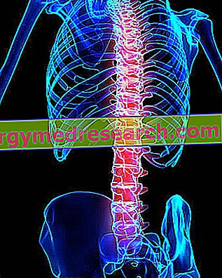
Generality
Tibia and fibula are the two long and even bones of the human body, which constitute the skeleton of the leg.

Bordering the femur, in a proximal position, and with the bones of the foot, in a distal position, the tibia and fibula are anatomically and physiologically important for their contribution to the knee and ankle joints.
Like all bones in the human body, tibia and fibula can also be broken.
What Tibia and Perone?
Tibia and fibula are the two equal bones of the human body, which constitute the skeleton of the leg . The leg is that anatomical portion of the lower limb including the upper thigh, and the foot, below (assuming, of course, to observe the human body in an upright position).
Anatomy
Tibia and fibula are two examples of long bones ; the peculiarity of the long bones is to be developed in length and to possess a narrow central portion, which takes the name of body or diaphysis, and two voluminous extremities, which are called proximal epiphysis and distal epiphysis .
Tibia and fibula run parallel to one another, with the tibia located in a medial position with respect to the fibula.
As leg bones, tibia and fibula adjoin the knee above and with the ankle below.
Brief review of the medial-lateral terms
Recalling that the sagittal plane is the anteroposterior division of the human body from which two equal and symmetrical halves are derived, " medial " means "near" or "closer" to the sagittal plane, while " lateral " means "far" or " farther "from the sagittal plane.
Examples:
- The second toe is lateral to the big toe, but is medial to the third toe.
- The ulna is medial with respect to the radius, which is lateral to the ulna (this is true if we assume that the upper limb is extended along the side and with the palm facing towards the observer).
Tibia
Between the tibia and the fibula, the tibia is the medial bone of the leg, as well as the most massive element of the skeleton of the latter.
NEXT EPIPHISTIC OF TIBIA
The proximal epiphysis of the tibia is the end of this bone of the leg closest to the femur, which is the bone of the anatomical region known as the thigh.
Obviously enlarged portion, the proximal epiphysis of the tibia owes its anatomical importance to its active participation in the knee joint.
Morphologically the proximal epiphysis of the tibia is distinguished:
- Two prominences called medial condyle and lateral condyle . The medial condyle resides on the inner side of the leg, while the lateral condyle is on the external side.

- The tibial plate . It is, in fact, the upper surface of the two aforementioned condyles.
At the center, the tibial plateau has two small pyramid-shaped bone processes, called intercondiloidean tubercles, which have the important task of anchoring the two meniscuses of the knee joint ; anteriorly, it has a rough depression, called anterior intercondylar fossa, on which the terminal head of the anterior cruciate ligament of the knee is attached; finally, posteriorly, it is provided with a second rough depression similar to the previous one, known as posterior intercondylar fossa, on which the terminal end of the posterior cruciate ligament of the knee is inserted.
- The tibial tuberosity . Located on the anterior surface of the tibia, just below the two condyles, it is a small bony relief that can be perceived by touch, which is still responsible for the terminal head of the patellar tendon .
The patellar tendon is a formation of fibrous tissue, which continues the tendons of the quadriceps femoral muscle and connects the knee patella to the tibia.
- The goose leg . Located at the same level as the radial tuberosity, but displaced on the medial surface, is another bony prominence, whose task is to anchor the terminal heads of the sartorius, gracilis and semitendinosus muscles .
Brief review of the proximal-distal terms
" Proximal " means "closer to the center of the body" or "closer to the point of origin"; " distal ", instead, means "farther from the center of the body" or "farther from the point of origin.
Examples:
- The femur is proximal to the tibia, which is distal to the femur.
- In the femur, the extremity bordering the trunk is the proximal end, while the extremity bordering the knee is the distal end.
BODY OF THE TIBIA
The body (or diaphysis) of the tibia is the portion of this bone of the leg interposed between the proximal epiphysis and the distal epiphysis.
On the body of the tibia, the following anatomical elements are relevant:
- The side surface . It is the portion of tibia designed to hook the so-called tibio-peroneal interosseous membrane . The interosseous tibio-peroneal membrane is a thin sheet of fibrous tissue which, interposed between the tibia and the fibula, indirectly unites the aforementioned bones of the leg.
- The back surface . It is the portion of tibia that, on a sort of bone crest called the soleus line, gives rise to the soleus muscle of the calf.
Did you know that ...
Between the tibia and the fibula, the tibia corresponds, in the upper limb, to the radium .
DISTAL EPIPHIST OF THE TIBIA
The distal epiphysis of the tibia is the end of this leg bone closest to the foot and farthest from the humerus.
Its anatomical relevance depends above all on its direct contribution to the ankle joint.
To distinguish the morphology of the distal epiphysis of the tibia are in particular:

- The lower margin . This, together with the inferior margin of the fibula, composes the region known as mortar ; the mortar is, in fact, a bone cavity, within which the talo (or astragalus ) takes place, which is one of the 7 bones that make up the tarsus of the foot .
- The medial malleolus . It is a bone process that develops in the inferior-medial direction, ie on the inside of the leg and downwards.
The function of the medial malleolus is to guarantee stability to the ankle joint.
- A groove in the posterior seat, within which the tendons of the posterior tibial muscle run .
- The fibular incisura . Based in a lateral position, it is a small shower recess that houses and hooks the distal end of the fibula.
| The joints that see the tibia involved: |
|
Fibula
Between the tibia and the fibula, the fibula is the lateral bone of the leg as well as the less voluminous skeletal structure.
Curiosity
In the sense in which the term "leg" indicates the anatomical tract between the hip and the ankle (and not, as in reality it would be more correct, the portion of the human body between the knee and the ankle), the expression "leg bones" includes not only the tibia and fibula, but also the femur.
PROXIMAL PERIPHYPE OF THE PERONE
Similar to an irregular square, the proximal epiphysis of the fibula is the end of this bone of the leg closest to the femur.
To characterize the morphology of the next epiphysis of the fibula are:
- A flattened surface in a medial position . Called facet, this surface serves to articulate the fibula to the tibia, precisely to the lateral tibial condyle.
- The styloid process (or apex ). With a medial position, the styloid process is a bony protrusion, which develops upwards and acts as an anchorage point for the terminal head of the quadriceps femoral muscle and for the terminal head of the lateral collateral ligament of the knee.

- A series of bone tubercles located on the anterior and posterior surfaces. Anteriorly, two tubercles emerge: on one the terminal head of the long peroneal muscle is inserted, while on the other one of the two ends of the anterior superior tibio-fibular ligament is attached (the other end is joined to the tibia).
At the rear, however, there is only one tubercle, which binds to itself one of the two ends of the so - called upper posterior tibio-fibular ligament (even in this situation, the other end is connected to the tibia).
The task of the aforementioned ligaments (anterior superior tibio-fibular and posterior superior tibio-fibular region) is to keep the tibia and fibula together.
Did you know that ...
Between the tibia and the fibula, the fibula corresponds, along the upper argon, to the ulna .
PERONE BODY
The body of the fibula is the portion of this bone of the leg situated between the proximal epiphysis and the distal epiphysis.
On the body of the fibula, anatomically speaking, 4 edges (antero-lateral, antero-medial, postero-lateral and postero-medial) and 4 surfaces (anterior, posterior, medial and lateral) stand out; among these anatomical elements, the most important element is, without doubt, the anterolateral border, as it is the anchor point, on the peroneal front, of the aforementioned interosseous tibio-fibular membrane.
DISTAL EPIPHIST OF THE PERONE
The distal fibular epiphysis is the extremity of this bone of the leg closest to the foot and ankle, and more distant from the femur.
The morphology of the distal fibular epiphysis is mainly characterized by:

- The peroneal malleolus (or lateral malleolus ). It is a bone process that extends on the lateral margin of the fibula and downwards, and which, together with the tibial malleolus (to which it resembles), contributes to the stabilization of the talus inside the mortar (remember that the astragalus is a of the 7 bones of the tarsus of the foot, while the mortar is the particular cavity of the inferior margin of the tibia delegated to receive the aforementioned tarsal bone).
- The articular facet that joins the fibula to the tibia at the level of their distal portions.
This facet occupies a medial position and is inserted in the aforementioned fibular incisura (recess of the tibia similar to the base of a shower).
| The joints in which the fibula participates: |
|
Ossification
Three ossification centers for each bone contribute to the definitive formation of tibia and fibula (therefore 6 in all).
For both the tibia and the fibula, the 3 ossification centers are located one where the body will be formed, one where the proximal epiphysis will come to life and one where the distal epiphysis will arise.
For the tibia, the first ossification center to activate is that of the body (VII week of fetal life), followed by that of the proximal epiphysis (shortly after birth) and that of the distal epiphysis (II year of life approximately) .
For the fibula, on the other hand, the activation of the ossification centers is activated first by that of the body (VIII week of fetal life), followed by that of the distal epiphysis (II year of life approximately) and by that of the proximal epiphysis (V year of life approximately).
Function
Tibia and fibula substantially cover 2 common functions, both equally important, which are:
- Form the knee joints (through contact with the femur) and ankle (through contact with the talus).
Knee and ankle two joints essential for locomotion; without them, in fact, the human being could not walk, run, jump, etc .;
- Inserting a series of muscles and ligaments, which allow the aforementioned joints to function properly and contribute to locomotion.
Exclusive Function of the Tibia
The tibia covers a third function, which is not the responsibility of the fibula. The function in question is to help support the weight of the body .
The fibula is excluded from supporting tasks of the body, as it is not in direct contact with the femur (this bone, therefore, does not weigh on the fibula).
diseases
Like practically all the other bones of the human body, also tibia and fibula can be subjected to fracture as a result of relevant trauma.
With regard to this topic, the simultaneous fracture of the tibia and fibula is particularly interesting; this is a very serious injury, with long healing times, which requires a period of immobilization and, often, also an ad hoc surgery.
The simultaneous fracture of the tibia and fibula concerns above all those who practice contact sports and those involved in serious road accidents .