Anatomy of skeletal muscle
Skeletal muscle is formed by a set of rather long, cylindrical cells with fusiform ends, called muscle fibers. If it is cut transversely it is noticed that these fibers are not isolated, but grouped in fascicles and wrapped by connective tissue. Elastic fibers, nerves and blood vessels run between one file and another, branching out to distribute to the various cells; the rich vascularization determines the typical coloring of the skeletal muscle (thanks to the myoglobline that circulates in the blood).
While the fleshy parts (muscular bellies) have a more or less intense red color, the tendinous parts have a pearly complexion.
The muscles are richly vascularized and innervated, and the course of the vessels and nerves is characteristic, always oblique and wavy to withstand the continuous changes in length to which each muscle goes during operation.
Muscle fibers are the largest cells in the body, although their dimensions are quite variable: from 10 to 100 µm as regards the diameter and between millimeter and 20 centimeters with regard to length. It is estimated that the human body contains about 250 million muscle fibers.
Muscle cells can hypertrophic, therefore they increase in size, but normally they cannot multiply. In other words, it is not possible to increase the number of fibers through training, but only the overall volume of those already existing.
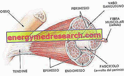
To recap: each muscle is formed by the union of several muscle bundles (or fragments); each bundle contains more fibers with a parallel course.
The size of the fascicles reflects the function of the muscle in question; for example, the muscles responsible for fine, tightly controlled movements have small fascicles and a relatively larger proportion of Perimysium (see below).
The entire muscle mass is covered by a sheath of fibro-elastic connective called epimysium, which has the task of containing it and protecting it during the execution of the movement itself. This sheath enters the muscular belly to constitute the perimysium and the endomysium: thus, each bundle is covered by a loose connective membrane called perimysium, while each single muscle cell is covered by a delicate connective membrane called endomysium.
- Epimysium or muscle band: sheath that covers the entire muscle
- Perimysium: sheath that covers the bundles of muscle fibers
- Endomysium: sheath that covers individual cells or muscle fibers
In the connective tissue interposed between the muscle fibers blood vessels and motor and sensitive nerve fibers run. Large vessels and nerves penetrate through the epimysium and divide to branch out through the muscle, in the perimysium and in the endomysium, reaching each individual fiber.
Anatomy of muscle fibers
When talking about muscles it is necessary to introduce a specific terminology. We have already seen how the cells that compose them are called fibers; the table shows the other terms to which we will refer in the rest of the article.
| Specific terminology related to muscles | |
| GENERIC TERM | MUSCULAR EQUIVALENT |
| Muscle cell | Muscle fiber or fibrocellula m. |
| Cell membrane | sarcolemma |
| Cytoplasm | sarcoplasma |
| Mitochondria | Sarcosomi |
| Endoplasmic reticulum | Sarcoplasmic reticulum |
The prefix sarc is derived from sarkos = meat.
Like the other cells of the body, the muscle fibers are surrounded by a plasma membrane, called sarcolemma; similarly, in analogy to the intracellular cytoplasm, this membrane contains the sarcoplasm.
First of all, inside the muscle cell we notice numerous nuclei. Every muscle fiber, in fact, derives from the union, during embryonic development, of multiple cells, called myoblasts, which fuse together. Therefore, muscle fiber is a syncytium (a term for multinucleated cells resulting from the fusion of multiple cells).
The nuclei of the muscle fibers are elongated, placed near the sarcolemma and particularly numerous, up to several hundred for each one. All this, with the aim of supporting the protein synthesis delegated, among other things, to the production of new contractile proteins (actin and myosin) to renew the worn ones.
Continuing our journey inside the muscle cell, we note that it is extraordinarily rich in voluminous mitochondria, arranged in parallel rows between the contractile elements; and it could not be otherwise. In fact, these organelles are responsible for the production of energy (ATP) necessary for muscle contraction.
Also in the cytoplasm, there is the presence of scattered granules of glycogen (a reserve energy substrate), lipid drops and myoglobin (a metalloprotein responsible for transporting and storing oxygen).
The sarcoplasm (ie the cytoplasm enclosed by the sarcolemma) is mainly occupied by:
- MITOCONDRI (energy production)
- LIPID DROPS (energy reserve)
- GLYCOGENO GRANULES (energy reserve)
- MIOGLOBINE (oxygen reserve)
- myofibrils and sarcoplasmic reticulum (illustrated in the next article)
Large and numerous mitochondria, glycogen granules and the presence of myoglobin ... a clear sign of the intense metabolic activity that takes place inside the muscle, with the aim of providing energy for contraction.