What is the Hypophyseal Adenoma
The pituitary adenoma is a benign tumor that develops from the cells of the pituitary gland, an endocrine gland responsible for the secretion of hormones that regulate numerous functions of the body. The clinical picture determined by a pituitary adenoma depends on many factors. Due to its large size, a macroadenoma can cause significant health effects due to the compression of neighboring structures (pituitary hypofunction, visual symptoms and neurological signs).

Diagnosis
History and visit of the patient
The first diagnostic approach is represented by the anamnesis and a careful objective examination . The doctor collects the information exposed by the patient concerning the symptoms and in particular the family medical history (presence in the family of other cases of pituitary cancer or some syndromes hereditary) .The physical examination allows to highlight symptoms and clinical signs characteristic of the disease and to re-evaluate the general state of health of the patient. The medical examination can include a neurological examination, to look for possible disorders of the nervous system, which could be caused by the compression exerted by the tumor mass.
Examination to evaluate visual acuity
The ocular clinical manifestations consist mainly of:
- Abnormalities in color vision (early symptom);
- Reduction of visual acuity (late symptom);
- Disorders of ocular motility (diplopia, ophthalmoplegia) or pupillary (mydriasis).
The ophthalmological evaluation allows to assess the vision, the visual field and diagnose any visual disturbances caused by a pituitary adenoma that compresses the optic chiasm. The patient undergoes an examination of the ocular fundus, to study the structures inside the eyeball, including the optic nerve. An additional determination consists in the campimetric examination, which allows to verify possible alterations at the level of the visual field: this test measures both the central vision (how much a person can see when he looks in front of him) and the peripheral one (how much a person can see in all other directions).
Laboratory investigations
When it is suspected that the pituitary adenoma has reduced the functionality of the healthy portion of the pituitary gland it is possible to resort to a simple blood test and a urine test . The laboratory investigations allow to evaluate the presence of possible hormonal alterations in the hypothalamic-pituitary axis and target organs, and allow to define if the adenoma determines hypopituitarism (pituitary insufficiency) or hypersecretory syndrome (overproduction with excess of one or more hormones).
Endocrine function tests include:
- basal dosage of pituitary tropins : these are tests that measure hormone levels in the blood. A higher or lower amount of these hormones produced by the pituitary gland can be a sign of pituitary adenoma. In particular, the levels of serum prolactin, TSH (thyroid-stimulating hormone), GH (growth hormone), ACTH (adrenocorticotropic hormone) and FSH (follicle-stimulating hormone) are measured.
- basal dosage of hormones produced by target organs : free T4 levels (FT4, free thyroxine), IGF-1 (insulin-1 growth factor), cortisolemia (serum cortisol dosage) and cortisoluria (urinary free cortisol) can be measured, 17β-estradiol (women) or testosterone (males).
Endocrinological assessments may also include inhibition and stimulation tests, which allow to evaluate the pituitary secretory reserve of certain hormones, possible dysfunctions in the hypothalamic stimulus, the hormonal response of target organs, etc.
Some of these investigations may include:
- ITT (Insulin Tolerance Test or insulin tolerance test);
- GH (growth hormone) stimulation test with arginine and GHRH;
- OGTT (Oral Glucose Tolerance Test or "oral glucose load" test);
- Cortisol dosage with ACTH stimulation;
- High dose and / or low dose suppression tests with dexamethasone.
Diagnostic imaging
Finally, to help the doctor define the position and the size of the pituitary adenoma, neuro-radiological tests are available, such as computerized tomography (CT) or magnetic resonance imaging (MRI) with contrast medium (generally, gadolinium). These techniques provide a series of detailed images of the internal structures of the brain and spinal cord, and allow the reliable identification of small lesions (from about 2 mm in diameter). The adenoma is highlighted as a hypodense mass in the pituitary parenchyma, with an intrasellar or extrasellar extension (with respect to the turgical saddle) and with alteration of the upper profile of the pituitary gland. This survey also highlights the degree of compression of the different structures adjacent to the tumor mass.
Care and Treatment
The pituitary adenoma therapy ideally involves the collaboration of various specialists (endocrinologist, neurosurgeon and neurologist) and is similar to that of other tumors:
- Drug therapy (generally, it is effective in tumors with prolactin or growth hormone hypersecretion, but not in those with ACTH hypersecretion);
- Radiotherapy ;
- Surgical removal of the tumor .
Early detection of pituitary adenomas is the key to successful treatment. Some factors influence the prognosis and the therapeutic options that can be adopted. The prognosis (probability of healing) depends on the type of tumor and whether it has spread or not to other areas of the central nervous system (brain and spinal cord) or to other parts of the body.
The treatment options of a pituitary adenoma depend on the following factors:
- Age of the patient and general health conditions;
- Type and size of the pituitary adenoma;
- Whether the tumor is a functioning adenoma that secretes hormones or not;
- If the tumor causes local disorders or other symptoms;
- If the tumor is spread to the surrounding structures adjacent to the pituitary gland or to other parts of the body;
- Whether pituitary adenoma has been diagnosed for a short time or tends to recur.
Pharmacological therapy
When the patient is suffering from a pituitary adenoma that overproduces a certain hormone, in some cases it is possible to resort to drug therapy. Often, the treatment involves the administration of inhibitory neurohormones ( dopaminergics and somatostatin analogues ), capable of limiting the secretion of excess hormones and reducing the size of the tumor mass.
The pituitary adenoma that responds best to this type of treatment is prolactinoma (pituitary secreting prolactin adenoma). Medical therapy often involves only the administration of dopaminergic agonists (they bind dopamine), which reduce prolactin secretion and potentially the tumor mass, thus allowing surgical removal to be avoided. From this point of view, it is important to consider that drug therapy must be placed after the differential diagnosis with macroprolactinoma, where the therapy is essentially surgical. The most widely used drugs for prolactin-secreting adenomas are bromocriptine and cabergoline : both are dopamine agonists that decrease prolactin secretion, alleviate symptoms and often reduce the size of the tumor mass. Possible side effects of these drugs include drowsiness, dizziness, nausea, vomiting, diarrhea or constipation, confusion and depression. While taking these drugs, some people may also experience compulsive behaviors.
Somatostatin analogues (octreotide, lanreotide, etc.) are available for the medical treatment of GH-secreting pituitary adenomas (growth hormone) and can also be used for some TSH-secreting adenomas . These drugs can have minor side effects, such as nausea, vomiting, diarrhea, stomach pain, dizziness, headache and pain at the injection site, although many of these tend to improve or disappear over time. They can also cause gallstones and can worsen diabetes if it has already been diagnosed in the patient.
Drug therapy plays an important role in the management of Cushing 's disease and in acromegaly .
If a pituitary adenoma causes a decrease in hormonal secretion or if surgical removal of the tumor has induced a deficit in hormone production, it may be necessary to resort to a specific replacement therapy to maintain hormone levels at normal values and to address pituitary insufficiency ( hypopituitarism).
Surgery
The treatment of large pituitary adenomas commonly consists of surgery. Usually, surgical removal is necessary when the pituitary adenoma compresses neighboring structures or is hypersecreting. The success of the surgery depends on the type of tumor, its location and size and whether or not the surrounding tissues are invaded. In most patients, surgical therapy allows a positive prognosis and complete recovery.
Surgery allows the complete removal of the pituitary adenoma and mainly involves two techniques:
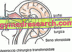
- Transsphenoidal approach . The localization of the pituitary allows a transsphenoidal intervention, where the surgeon uses endoscopes to access the sphenoid bone, passing through the nasal cavity or under the upper lip. This procedure is minimally invasive, does not involve external incisions, minimizes complications and hospitalization time. However, the transsphenoidal intervention allows to treat only adenomas of small size (microadenomas) and with a low degree of invasiveness.
- Transcranial approach (craniotomy) . Some macroadenomas extend into the brain cavity and may require the opening of the skull, through an incision in the scalp, to access the tumor. Often, the procedure is associated with drug therapy and post-operative radiotherapy.
Radiotherapy
Some pituitary adenomas cannot be removed surgically, as they are not easily accessible, while others may be refractory to treatment with drugs. Radiation therapy uses high-energy radiation, which selectively acts on the target tumor (in general, the surrounding brain structures receive only a fraction of the radiation). Among the different methods we mention conventional and stereotactic radiotherapy (gamma-knife).
Radiation therapy can be effective in controlling the growth of pituitary adenomas or in destroying any residual tumor cells (post-operative radiotherapy). However, treatment with radiation may, in some cases, result in pituitary insufficiency, which generally occurs several years after treatment and necessitates hormone replacement therapy.
Prognosis and life expectancy
The prognosis of pituitary adenomas is positive: surgical excision is safe and allows to restore normal hormonal production. Remission (complete healing) can be achieved in 90% of patients with microadenomas and in about 50-60% of macroadenomas. Furthermore, pituitary adenoma is a type of tumor that tends to hardly recur. In some cases, pituitary insufficiency may appear after surgery: this condition is a rare occurrence in microadenomas, whereas it is more frequent in macroadenomas (30% of cases).


