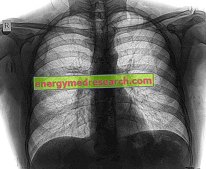
The chest X -ray, also known as chest radiograph, is a widely practiced diagnostic test, which has the purpose of reproducing the organs and bone structures of an individual's chest on a photographic plate (or a digital detector).
Generally, it is performed when an individual suffers from dyspnea and possibly even severe and / or persistent cough, chest pain, chest pain due to trauma, fever.
Thanks to the reproductions that a chest X-ray can provide, doctors can analyze:
The lungs . The RX-thorax allows diagnosing various morbid conditions, including: pulmonary infections, cystic fibrosis, lung carcinomas, pulmonary emphysema, pneumothorax, etc.
The heart . Possible abnormalities or cardiac malformations can be identified, such as valvular defects or a condition called cardiac tamponade.
The blood vessels that depart from the heart . The defects of the vessels that connect the heart to the lungs or the vessels that connect the heart to the various districts of the body (aorta) can be seen.
The presence of calcium deposits in the blood vessels .
The presence of bone fractures .
Changes occurred at cardiac or pulmonary level after surgery.