What is the Optic Nerve?
The optic nerve represents the beginning of the optical pathways, that is, the set of structures that, starting from the retina, connect the eyeball to the brain.
This component is indispensable for correctly activating the vision. The optic nerve is in fact responsible for the transfer of the electrical impulses resulting from the retinal receptor transduction, thus allowing visual perception.
Structure
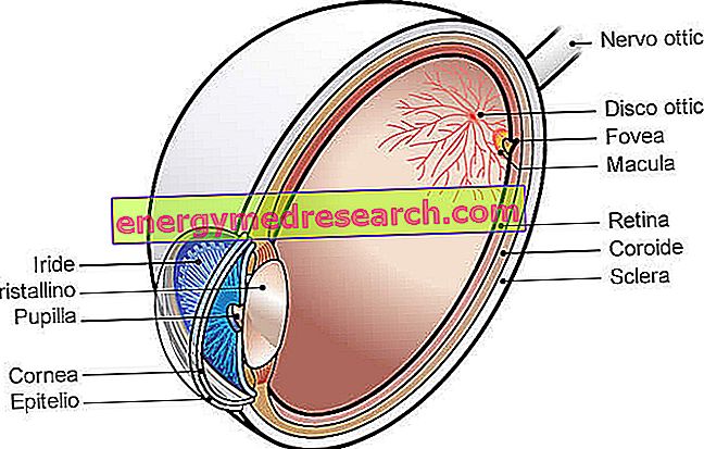
The optic nerve represents the second pair of cranial nerves; originates from the confluence of retinal optical fibers at the optic papilla (also called optic nerve head ).
Its structure is comparable to an electric cable that has many copper wires inside (more than 1, 200, 000 nerve fibers divided into about 200 bundles). Each single fiber (similar to a thread) corresponds to a small area of the retina, so each bundle coincides with a more extensive retinal region. Despite the partial crossing of the nerve fibers that occurs at the level of the optic chiasm, this arrangement is maintained up to the visual cortex.
The course of the optic nerve can be divided into four segments:
- Intraocular segment (very short portion that begins in the eyeball at the level of the optic disc, then crosses the choroid and the cribrosum diaphragm of the scleral canal to escape from the eye);
- Intraorbital segment (continuous in the orbit, that is from the posterior pole of the eye up to the optical channel of the sphenoid bone; it is the longest portion - about 2.5 cm - of the optic nerve);
- Intracanalicular segment (short section included in the optical channel);
- Intracranial segment (extends from the middle cranial fossa to the optic chiasm).
Like the white substance of the brain, the optic nerve has a support network made up of astrocytes, microglia and oligodendrocytes.
Unlike the other cranial nerves which have a thin sheath called neurilemma (consisting of Schwann cells), the axons of the optic nerve are lined with myelin produced by oligodendrocytes.
For this reason, the optic nerve is considered as part of the central nervous system .
Note : having no neurilemma, the nerve fibers that make up the optic nerve have very little regeneration capacity. Therefore, any damage is irreversible and can lead to blindness.
Even the white encephalic substance has the same characteristic.
Similarly to the brain, the optic nerve is enveloped by the meninges (dura mater, arachnoid and pia mater) and has a minimal amount of CSF (between the pia mater and the arachnoid). This explains its susceptibility to being involved during meningitis.
Furthermore, presenting common characteristics with the white encephalic substance, the optic nerve is particularly vulnerable to demyelinating diseases (multiple sclerosis) and encephalitis.
Retina and origin of the optic nerve
The retina is the photosensitive surface of the eye, formed by:
- Cones and rods : photoreceptor cells placed in the most superficial retinal layer and deputed to convert images into electrical signals (phototransduction), which are transmitted to the brain via the two optic nerves. Cones and rods, when exposed to light or darkness, in fact, undergo conformational changes, which modulate the release of neurotransmitters. These perform an excitatory or inhibitory action on the bipolar cells of the retina.
- Bipolar cells : they are connected on one side to the photoreceptors and on the other to the ganglion cells of the innermost layer, whose axons give rise to the optic nerve. Bipolar cells are capable of transmitting graduated potentials.
- Ganglion cells : their axons form a bundle that converges on the optic disk and exits from the eyeball, proceeding towards the diencephalon as an optic nerve (II pair of cranial nerves); in response to retinal receptor transduction, ganglion cells generate action potentials aimed at the central nervous system.
In other words, the optic nerve is the extension of the nerve endings of the photoreceptors of the retina.
Note. Each cone, like each rod, controls a specific receptor field. Each image is, therefore, the result of the elaboration of information provided by the entire receptor population. A significant amount of processing already takes place at the level of the retina, thanks to the interactions between the different cell types, before the information is sent to the brain.
Optical disk
The optic disc (or optic papilla) represents the onset of the optic nerve. On examination of the ocular fundus, this area of the retinal plane appears as a small oval area of markedly white color, since it is made up of myelinated axons about to leave the eyeball.
The optic disc is located below and medially to the posterior pole of the eye, at a distance of about 4 millimeters from the macula.
From the center of the optic disc, the blood vessels that supply the eye emerge.
Blind spot
Near the optic disc, there is the blind spot, defined thus for the lack of photoreceptors and other retinal cells. The light that reaches this area goes completely unnoticed and cannot generate electrical impulses, however in the visual field an empty area is not perceived. In fact, involuntary eye movements keep the image moving and allow the brain to fill in the missing information.
How to demonstrate the presence of the blind spot
A simple experiment can demonstrate the presence of the blind spot:
- On a white sheet draw a + sign on the left and a sign - on the right, respecting a distance of 5 cm from each other.
- Cover the right eye and observe the sign - with the left eye.
- Position the sheet at a distance of about 30 cm and fix the mark with your left eye, keeping your gaze fixed on the image.
- Moving the head back and forth, it should be noted that the + sign disappears and reappears alternately from one's sight. This happens because the reflected light of the + sign hits the optical disk, so it cannot be perceived.
Optical pathways

- Optic nerve;
- Optic chiasm;
- Optical tract;
- Lateral geniculate nucleus (or body);
- Optical Gratiolet radiation (projection fibers).
After about five centimeters from the optical foramina, the optic nerves coming from the two eyes reach the base of the brain in front of the trunk of the brain, to form the optic chiasm . As anticipated, at this level, there is a partial crossing: about half of the fibers from each eye proceed towards the lateral geniculate nucleus of the ipsilateral thalamus, while the other half reaches the lateral geniculate nucleus of the opposite side. Consequently, each cerebral hemisphere receives visual information from the lateral half of the ipsilateral retina and from the medial half of the contralateral retina. Therefore, both eyes receive information from both visual fields.
After the optic chiasm, ganglion cell axons travel in a bundle of fibers called the optic tract, which terminates in the lateral geniculate nucleus.
The lateral geniculate nuclei act as processing centers that send visual information to the reflex centers of the brainstem and to the cerebral cortex. For example, pupillary reflexes and reflexes that control eye movements are triggered by information from the lateral geniculate nuclei. At this level, the optic tract forms synapses with neurons that reach the visual cortex of the occipital lobe ( Gratiolet optical radiation ), where the formation of visual sensation occurs.
What is the optic chiasm for?
The partial crossing of the nerve fibers that occurs at the level of the optic chiasm allows the visual cortex to receive a composite image of the entire visual field.
Each eye, in fact, receives a very different image because:
- The fovee (central portions of the macula delegated to the finest vision) are placed at a certain distance;
- The nose and orbit block the vision of the opposite side.
The areas of cortical association and integration therefore compare the two perspectives and use them for deep perception, in order to obtain a complete image of the entire visual field.
Functions
The function of the optic nerve is to transmit nerve impulses generated at the level of the retina to the brain.
In this way, this component of the visual system allows the interpretation of the signals perceived in the images that we actually see when we open our eyes.
Diseases of the optic nerve
There are many pathologies that can involve the optic nerve. In fact, optical neuropathies of metabolic, infectious, degenerative (multiple sclerosis), infiltrative (eg sarcoidosis), autoimmune, vascular (ischemic and aneurysmal compressions), toxic-deficient, inflammatory, neoplastic, traumatic and drug-induced neuropathic are recognized.
Furthermore, congenital malformations are possible, such as coloboma, Leber's optic atrophy and optic nerve aplasia.
Symptoms
Damage or compression of the optic nerve results, symptomatically, in defects of the visual field (such as scotomas and hemianopsia), alteration of the pupillary reflex and decrease in visual acuity of various degrees. Pain may also occur in the back of the eye (especially when moving the globe), headache and altered color perception (reduced or staggered).
If the suffering of the optic nerve is chronic, therefore prolonged over time, it can also lead to atrophy. Glaucoma in the terminal phase is characterized by this sign.
Optic neuritis
Optic neuritis is an inflammation of the optic nerve that recognizes several causes. In fact, it can be associated with infectious diseases (such as sinusitis and meningitis) and autoimmune diseases (optic neuromyelitis).
Often, optic neuritis is the onset symptom of multiple sclerosis (demyelinating pathology that affects portions of the central nervous system) and is commonly present in the phases of exacerbation of the disease.
Inflammation of the optic nerve may also result from systemic diseases (such as systemic lupus erythematosus, connective tissue diseases, etc.) and neoplastic diseases. The total or partial infarction of the optic papilla and alcohol and tobacco intoxication (which influence the correct absorption of nutrients essential for the correct functioning of the nervous system) can also involve neurological pain affecting the optic nerve.
There are also isolated forms in which a specific cause cannot be established.
Optic neuritis involves visual disturbances such as the loss of part of the visual field and diplopia.
papilledema
The papilledema (or edema of the papilla) is the swelling of the optic disc on the retinal plane. This pathological condition can be caused by an increase in intracranial pressure, secondary, for example, to tumors, meningitis, head injuries and bleeding.
In other cases, edema is a consequence of glaucoma: intraocular hypertension involves a typical aspect of the optic papilla, which increases its excavation in relation to the progression of the pathology.