Generality
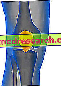
The patella, or patella, is the protruding bone, detectable by touch, which is located on the front of the knee joint.
Fundamental to the extension of the leg, this bone element is very reminiscent of an inverted triangle.

In addition to guaranteeing the extension of the leg (fundamental mechanism for locomotion), the patella also serves to increase the efficiency of the quadriceps muscle and to protect the internal anatomical structures of the knee.
What is the Rotula
The patella, or patella, is the protruding and palpable bone, which resides in front of the knee joint, at the border between the thigh bone (the femur) and the leg bone (the tibia).
IT IS A SESAMOID BONE
Anatomy experts include the patella in the sesamoid bone category, ie those bones that have a similar appearance to sesame seeds.
Other examples of sesamoid bones, present in the human body, are: the pisiform bone of the hand carpus and the lenticular process of the anvil (one of the three ossicles of the middle ear).
Anatomy
The patella is a thick, triangular shaped bone element, with an important front surface and an equally important posterior surface.
As a triangle, it is inverted: the apex of the patella is projected downwards, therefore from the side of the tibia; while the base is facing upwards, therefore it is in the direction of the femur.
Important note: meaning of medial and lateral
Medial and lateral are two terms with the opposite meaning. However, to fully understand what they mean, it is necessary to take a step back and review the concept of the sagittal plan.
The sagittal plane, or median plane of symmetry, is the antero-posterior division of the body, a division from which two equal and symmetrical halves are derived: the right half and the left half. For example, from a sagittal plane of the head derive a half, which includes the right eye, the right ear, the right nasal nostril and so on, and a half, which includes the left eye, the left ear, the left nasal nostril etc.
Returning to the medial-lateral concepts, the word media indicates a relationship of proximity to the sagittal plane; while the word side indicates a relationship of distance from the sagittal plane.
All anatomical organs can be medial or lateral with respect to a reference point. A couple of examples clarify this statement:
First example. If the reference point is the eye, it is lateral to the nasal nostril, but medial to the ear;
Second example. If the reference point is the second toe, this element is lateral to the first toe (toe), but medial to all the others.
BOND WITH THE FEMORE
The patella is in communication with the femur by means of a set of tendons, which belong to the muscles that make up the so-called quadriceps . Based in the thigh, the muscles that make up the quadriceps are: the rectus femoris, the intermediate broad muscle, the vastus medialis muscle and the lateral vastus muscle.
The tendons of the rectus femoris and the vast intermediate attach to the base of the triangle that ideally represents the patella; the tendons of the vastus medialis and the vastus lateralis, on the other hand, join, respectively, to the medial border (internal) and to the lateral (external) edge of the patella.
What is a tendon? Difference from a ligament
A tendon is a formation of fibrous connective tissue that joins a muscle to a bone element.
A ligament is a very similar structure, but with the substantial difference that unites two distinct bones or two different parts of the same bone.

Figure: quadriceps muscles and their tendon insertions on the patella. The intermediate broad muscle resides under the rectus femoris and this explains why the image shows the quadriceps at a higher level and at a deep level.
BOND WITH THE TIBIA
The patella communicates with the tibia through a ligament, called the patellar ligament (or patellar ).
Welded at the apex of the triangle that ideally represents the patella, the patellar ligament joins the tibia in a bony prominence of the proximal end, bony prominence that doctors define with the term tibial tuberosity .
Many readers have probably heard of patellar tendon instead of a patellar ligament. The two terms, at this juncture, are equivalent (thus identifying the same anatomical element), for a very specific reason: the structure of connective tissue, which joins the patella to the tibia, is a continuity of the tendons of the quadriceps muscles.
REAR SURFACE

The posterior surface of the patella has two areas with a particular appearance: the medial facet and the lateral facet . Together, these two areas serve to articulate the patella at the distal end of the femur (NB: by distal end of the femur, we mean the bone section most distant from the trunk).
Entering more specifically:
- The medial facet articulates with the medial condyle of the femur ;
- The lateral facet articulates with the lateral condyle of the femur .
What are the medial and lateral femoral condyles?
The medial condyle and the lateral condyle of the femur are two oblong and rounded prominences, which are located at the end of the femur.
Their postero-inferior surface is articulated with the tibia and the medial and lateral meniscus of the knee, while their anterior surface articulates with the patella.
MEASUREMENTS OF THE ROTULA OF AN ADULT PERSON

Figure: the femur and its main sections: proximal end, body and distal end. The reader may note the presence of the medial and lateral ones at the level of the distal end.
In adults, the patella has a surface extension of about 12 cm2.
Moreover, all around, it has a cartilage coating with a thickness not exceeding 6 millimeters.
Functions
The patella performs two important functions:
- It allows the extension of the leg, a fundamental movement for locomotion.
- Optimize the "work" done by the quadriceps muscles . According to experts, the tendon insertions of the quadriceps on the patella act as a lever. This leverage function allows the connected muscles (rectus femoris, broad intermediate etc.) to increase their "work" force, during a movement, a run, etc.
- It guarantees protection to the articular elements that make up the knee . Inside the knee there are some very fragile structures, such as the anterior and posterior cruciate ligaments. The rupture of these ligaments (in particular the anterior cruciate ligament) requires ad hoc surgery, as their repair is almost impossible.
Diseases of the patella
The most known problems that can affect the patella are: dislocation (or dislocation), rupture of the patellar tendon and fracture.
LUXURY OF THE ROTULA
The dislocation of the patella, or patellar dislocation, is when the patella slips out of its normal position.
Due to significant knee pain, this condition usually arises after a considerable impact on the patella or due to a sudden knee twist.
In general, the patella performs a lateral slide; however, it could also slip in a medial direction (ie inward).
According to some statistical researches, the patella dislocation represents 3% of knee injuries and mainly affects subjects who frequently participate in contact sports, such as rugby, football, American football and ice hockey.
BREAKAGE OF ROTULEO CURTAINS
The rupture of the patellar tendon (or patellar ligament) is an injury that affects the formation of fibrous tissue that connects the patella to the tibia.
The individuals who practice high-level contact sports, such as football, rugby, etc. suffer the most.
The only therapeutic solution, adoptable in the presence of a rupture of the patellar tendon, is the surgical reconstruction of the aforementioned tendon.
ROUTE FRACTURE
The patella can fracture like all other bones in the human body.
In most cases, fractures of the patella have a traumatic origin. However, they can also be the result of a sudden and unnatural contraction of the quadriceps.
Generally, from the breakage of a patella originate two bone fragments: one in an upper position (also called proximal), which is connected to the quadriceps tendons, and one in a lower position (also called distal), which is connected to the patellar tendon.
The patella fracture is very common in males between 20 and 50 years.


