Generality
Carotids are two large arterial vessels in the neck, whose branches supply the central nervous system and facial structures.
A right carotid artery and a left carotid artery are distinguished, respectively. Like the vertebral arteries, they have the function of bringing blood to the brain. In addition to oxygenating the cerebral districts, the carotid arterial system also deals with spraying the areas of the head corresponding to face and eyes. The most common pathologies that compromise the carotid function are arteriosclerosis and atherosclerosis.
- Arteriosclerosis causes a loss of elasticity and contractility, as well as a modification of the vessel caliber.
- Atherosclerosis causes the formation of plaques (atheromas) that occlude the lumen of the arterial vessel.
Anatomical review of arteries
Arteries are vessels that originate directly or indirectly from the heart and, receiving oxygenated blood from the latter, supply all the tissues and organs of the human body. The blood in the arteries flows in the centrifugal direction, that is towards the periphery.
As you move away from the heart, the arterial system gradually branches. The caliber of the vessels is therefore reduced; in this regard, we can distinguish:
- Large-caliber vessels, whose diameter measures at least 7 mm. They are the arteries that originate from the heart, like the aorta or the carotids themselves
- Medium caliber vessels, whose diameter measures between 7 mm and 2.5 mm.
- Small-caliber vessels, whose diameter measures less than 2.5 mm.
- Arterioles, the last branches of the arterial system. They measure less than 100 microns.
As for the veins, also the wall of the arteries consists of 3 concentric layers, of variable thickness and structure depending on the size of the vessel. The 3 layers are:
- The intimate, endothelium-coated cassock . It is the most internal part of the vase.
- The average habit, made up of elastic and muscular fibers. The elastic component prevails in large vessels; while the muscular component prevails in medium caliber vessels
- The adventitious cassock, consisting of connective tissue and, sometimes, muscle and elastic fibers. It is the outermost part of the vase.
Anatomy of the carotid arteries
Carotids are classified as large-caliber arteries, as they originate from the heart. They spray the following districts or areas of the head:
- Brain.
- Face.
- Eyes.
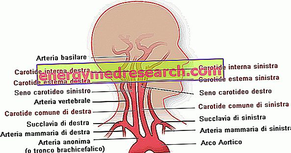
The carotid arteries are two, right and left, and each has two terminal branches, called the external carotid artery and internal carotid artery. Therefore, the carotid arterial system can be schematized as follows:
- Two common carotid arteries, right and left.
- Two branches for a single common carotid artery:
- the external carotid artery
- the internal carotid artery .
The right common carotid arises from the anonymous aorta, or brachycephalic, right, one of the first vessels that arise from the arch of the aorta. The common left carotid arises directly from the arch of the aorta. Their length is clearly different: the right is shorter.
The two vessels, right and left, go upwards and end about one centimeter above the upper portion of cartilage that makes up the thyroid. Here they each divide into two branches, the external carotid artery and the internal carotid artery.
Originating directly from the aortic arch, the left carotid artery establishes relationships with other districts of the body, adjacent to it, at the endothorax level. It relates to:
- The anonymous vein on the left, front.
- The trachea and the esophagus, behind.
- The vagus nerve on the left sideways.
In the neck, the two common carotids, right and left, contract the same relationships with neighboring organs. They contact:
- The internal jugular vein and with the vagus nerve of each side. All together, they form the neurovascular bundle of the neck .
- Pharynx, esophagus, larynx, trachea, thyroid gland and nerves are the medial relationships.
The external carotid artery crosses different muscles (digastric and stylohyoid), venous vessels (tirolinguofacial) and nerves (hypoglossus) of the head, reaching the parotid gland.
Proceeding from the bottom upwards, the external carotid artery emits the following side branches:
- Upper thyroid artery.
- Lingual artery.
- Sternocleidomastoid artery.
- External maxillary artery.
- Occipital artery.
- Pharyngomeningeal artery.
- Posterior auricular artery.
- Parotid arteries.
Finally, it ends at the level of the jaw. Here it branches into:
- Superficial temporal artery.
- Internal maxillary artery.
The internal carotid artery, on the other hand, ends inside the skull. It also contracts with muscles, venous vessels and nerves of the head. It has numerous reports, the main ones established with:
- The digastric, stylohyoid, pharyngeal and styloglossus muscles
- The internal jugular vein
- Vagus nerve, glossopharyngeal nerve and hypoglossal nerve.
The internal carotid artery, in its terminal point, pierces the dura mater and penetrates into the endocranium (the inner wall of the skull). In this area, it makes contact with various nerves of the eye.
The collateral ramifications are the following:
- Caroticotympanic artery
- Ophthalmic artery
- Middle cerebral artery
- Anterior corioid artery
- Rear communicating artery.
The terminal branch, on the other hand, is the anterior cerebral artery.
diseases
The most common pathology affecting the carotid system is arteriosclerosis . It is a typical disease of the arteries and shows the following characteristics:
- Increase in consistency, followed by tissue hardening of the vessel wall. In this case, we are talking about sclerosis .
- Modified vessel thickness: thickening or thinning.
- Modified vessel length: the artery lengthens and becomes more tortuous.
- Internal surface modified: it becomes irregular.
- Modified caliber: dilation or stenosis of the vessel.
These characteristics determine two typical consequences of arteriosclerosis:
- Decreased vessel elasticity.
- Decrease in vessel contractility.
Spraying through the arteriosclerotic vessels is therefore insufficient and generates serious complications in inadequately oxygenated tissues. This is what happens to the carotid system: brain districts, the face and eyes lose their normal capacity. The effects, unfortunately, are not limited to these sites: in fact, there is also a loss of control of the limbs innervated by the brain areas no longer reached by a correct blood flow.
Among the forms of arteriosclerosis, various pathologies are included from particular clinical pictures. One of these is atherosclerosis . The other pathological forms affect medium and small caliber arteries, therefore, this is not the right place to talk about them.
Atherosclerosis is a disease typical of the most elastic arteries present in the human body: therefore, it affects, preferably, the large-caliber arterial vessels, which originate from the heart; secondly, it also affects the medium caliber vessels that originate from the upper caliber arteries.
Atherosclerosis has the following general characteristics:
- The medium cassock (in the innermost layers), and, above all, the intimate habit are characterized by the presence of focal plates, forming reliefs and made up of fibrolipid material. These plaques are called atheromas . Their distribution is, therefore, well localized.
- The fibrolipid consistency of the atheromas is a consequence of an accumulation of lipid material and the proliferation of the fibrous component of the connective tissue.
- The atheromas can be distributed as foci, but never as continuous structures that affect the arterial vessel: the atherosclerotic artery always presents free areas.
- It has a slow and progressive evolution over time.
- It affects every individual, with greater incidence in the male. The first processes of atherosclerosis can develop already between the 2nd or 3rd decade of life. Around the 6th decade of life, atheromatous lesions are common and obvious.
- It can be asymptomatic.
- Complications: myocardial infarction, intestinal infarction, cerebral hemorrhage, aneurysms and senile gangrene of the lower extremities.
In the carotids, the atheromatous plaques are distributed in a variable way and often become the site of thrombotic deposits, obstructing the lumen. This pathological situation is known as carotid stenosis .
Finally, other pathologies affecting the carotid artery are due to trauma, aneurysms and obliterating thromboangioitis .