Generality
The femoral artery is the large even arterial vessel that crosses the thigh and which, with its branches, provides blood circulation to numerous districts of each lower limb.
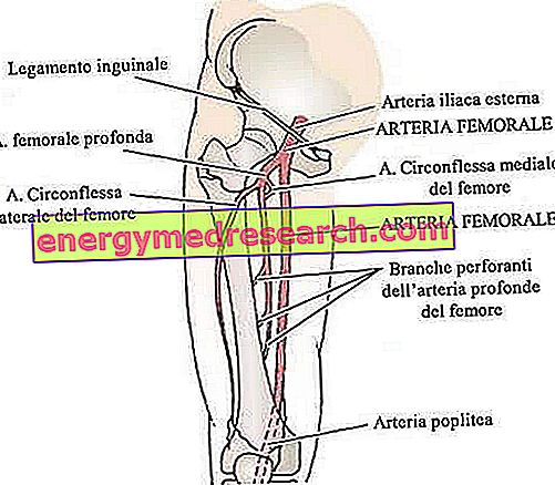
The femoral artery starts behind the inguinal ligament (beginning of the thigh) and ends where the popliteal artery begins, almost at knee height. Along its path along the thigh, it gives rise to numerous branches, including: the superficial iliac circumflex artery, the superficial epigastric artery, the superficial external pudendal artery, the deep external pudendal artery and the geniculate descending artery.
Anatomically speaking, the femoral artery can be divided into three parts: the common femoral artery, which is the very first tract, the deep femoral artery, which is the section following the common femoral artery, is the superficial femoral artery, which is the terminal trait.
What is the femoral artery?
The femoral artery is a large arterial vessel of equal size, which crosses the region of the thigh and gives rise to various branches (or branches), deputed to the blood supply of numerous districts of the lower limb .
In anatomy books, the femoral artery is described as "the main arterial vessel in the lower limbs of the human body".
ORIGIN OF THE NAME
The femoral artery is so called because of the close relationship that the arterial vessel in question establishes with the femur, ie the thigh bone.
Anatomy
The femoral artery begins approximately at the height of where the thigh begins. The precise starting point is behind the inguinal ligament . Here, the femoral artery passes through an anatomical area, called the femoral triangle (or Scarpa's triangle ).
The femoral triangle derives from the particular arrangement of some thigh muscles, including the sartorius muscle, the long adductor muscle, the pectineus muscle and the ileopsoas muscle; in addition to the femoral artery, it contains the femoral vein, the femoral nerve, the femoral sheath, the inguinal lymphatic vessels and the deep inguinal lymph nodes.
Continuation of the so-called external iliac artery, the femoral artery makes a path along the thigh, through which the anatomists divide it into three parts:
- The common femoral artery . It is the very first stretch of the femoral artery;
- The deep femoral artery . It is the section following the common femoral artery;
- The superficial femoral artery . It is the last stretch of the femoral artery; concludes his path giving rise to the popliteal artery, deputed, with its branches, to the blood circulation of the knee, leg and foot.
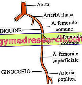
Figure: parts of the femoral artery. The design ends at the level of the popliteal artery, but it should be pointed out that, from the knee down, an articulated network of other arterial blood vessels comes to life.
REPORTS OF FEMORAL ARTERY
In its path along the thigh, the femoral artery makes contact with various anatomical structures.
The complete picture of the relations established by the femoral artery is the following:
- Anteriorly (ie in front of the femoral artery): in its very first section, the femoral artery is located very far on the surface, just below the skin. Later, it goes deeper and deeper, until it passes below the sartorius muscle (which, therefore, is in front of it).
- Posteriorly (ie behind the femoral artery): behind the femoral artery takes place the large psoas muscle, which separates it from the hip joint, from the pectineus muscle and from the long adductor muscle.
- Medially (ie in a medial position with respect to the femoral artery): on the medial side, the femoral artery is bordered by the femoral vein.
- Laterally (ie laterally with respect to the femoral artery): on the lateral side, the femoral artery is bordered by the femoral nerve and its branches.
In anatomy, medial and lateral are two terms of opposite meaning, which serve to indicate the distance of an anatomical element from the sagittal plane .
The sagittal plane is the anteroposterior division of the human body, from which two equal and symmetrical halves are derived.
Mediale means "near" or "closer" to the sagittal plane, while lateral means "far or" farther "from the sagittal plane.
BRANCHE OF FEMORAL ARTERY
The femoral artery gives rise to numerous branches. These branches are:
- The superficial iliac circumflex artery . It is the smallest branch of the femoral artery; it rises near the superficial epigastric artery (another branch of the femoral artery), it runs parallel to the inguinal ligament and ends in the region where the anterior superior iliac spine takes shape.
- The superficial epigastric artery . It is a branch of very small size, which, proceeding upwards, crosses the inguinal ligament and ends up in the umbilical region, where it supplies blood to some of the tissues present here.
- The superficial external pudendal artery . It is another branch of small size, which, proceeding in a medial direction with respect to the femoral artery, supplies blood to the skin of the scrotum, in man, and to the labia, in the woman.
- The deep external pudendal artery . It is a branch similar in terms of course and function to the previous one. Along its path, it passes through the pectineus muscle and the long adductor muscle; it is covered by the so-called band lata.
- The geniculate descending artery . It is a small branch that originates almost at the end of the femoral artery, before the latter becomes a popliteal artery. His job is to supply the knee joint with blood.
Some books on human anatomy also consider the femoral artery as a branch of the femoral artery. Here, even to simplify understanding, the deep femoral artery is simply considered a tract of the femoral artery.
The deep femoral artery starts about 4 centimeters lower than the inguinal ligament. Flowing between the pectoral muscle and the long adductor, it gives rise to several sub-branches: the medial circumflex artery of the femur, the lateral circumflex artery of the femur and the three perforating arteries.
Functions
As anticipated in the definition of the femoral artery, the latter is the main blood vessel of each lower limb of the human body. In fact, also thanks to its branches, it deals with supplying numerous anatomical structures present in the thigh and in the immediate vicinity of the thigh.
clinic
The initial tract of the femoral artery lies just below the skin. This makes the arterial vessel in question a convenient point of entry for catheters, during diagnostic and / or therapeutic procedures that have as their object the heart, the brain, the kidneys, the arms or the legs.
FEMORAL AND FEMORAL ARTERY
Once again thanks to its very superficial position in the first tract, the femoral artery is an arterial blood vessel that allows the measurement of systolic blood pressure . The systolic blood pressure measurement performed by palpation of the femoral artery is called the " femoral pulse ". The "femoral pulse" allows to detect systolic blood pressures greater than 50 mmHg.
Associated pathologies
The femoral artery is an arterial vessel that is very susceptible to a medical condition known as peripheral arterial disease .
Peripheral arteriopathy is a fairly common problem, characterized by the accumulation of fatty deposits inside an artery and the narrowing of the latter, precisely following the accumulation of the aforementioned adipose deposits. The narrowing of an artery affects the ability of the same to make the blood flow inside it.
People with severe peripheral arterial disease of the femoral artery need surgical treatment to restore normal blood flow.