Generality
The scapula is the even bone, located posterolateral to the thoracic cage, which articulates the trunk at the upper limb of each side of the human body.

The shoulder blades have two main functions: hooking the head of the humerus to their glenoid cavity, thus forming the so-called glenohumeral joint (or shoulder joint), and inserting it into the initial ends of the muscles constituting the rotator cuff.
Like any bone in the human skeleton, the scapula can fracture. Fractures of the scapula are very rare events, which generally result from severe trauma to the chest.
What is the Scapula
The scapula is the even bone, posteriorly posterior to the rib cage, which connects the trunk to the upper limb of each side of the body.
The junction between the trunk and upper limb (arm) is the so - called glenohumeral joint .
The glenohumeral joint - better known as the shoulder joint - involves the lateral margin of the scapula and the head of the humerus . The humerus is the arm bone and the head of the humerus is a bone projection with medial orientation.
The scapula is a flat triangular bone, which occupies a certain position by virtue of its union with the clavicle. On it there are numerous muscles of the upper shoulder-limb anatomical tract.
Brief review of the concepts: sagittal plane, medial position and lateral position
In anatomy, medial and lateral are two terms with the opposite meaning. However, to fully understand what they mean, it is necessary to take a step back and review the concept of the sagittal plan.

Figure: the plans with which the anatomists dissect the human body. In the image, in particular, the sagittal plane is highlighted.
The sagittal plane, or median plane of symmetry, is the antero-posterior division of the body, a division from which two equal and symmetrical halves are derived: the right half and the left half. For example, from a sagittal plane of the head derive a half, which includes the right eye, the right ear, the right nasal nostril and so on, and a half, which includes the left eye, the left ear, the left nasal nostril etc.
Returning to the medial-lateral concepts, the word media indicates a relationship of proximity to the sagittal plane; while the word side indicates a relationship of distance from the sagittal plane.
All anatomical organs can be medial or lateral with respect to a reference point. A couple of examples clarify this statement:
First example. If the reference point is the eye, it is lateral to the nasal nostril of the same side, but medial to the ear.
Second example. If the reference point is the second toe, this element is lateral to the first toe (toe), but medial to all the others.
SYNONYM OF SCAPOLA
As an alternative to the word scapula, doctors use - even if, in truth, very rarely - the synonym homoplated . Omoplata derives from the union of two Greek terms: ὦμος, which means "shoulder", and πλατύς, which means "broad".
Anatomy
The scapula has a supporting region, called the body, on which various bony projections and other particularities develop, such as more or less short ridges, pits, convexity, etc.
To simplify the study of the scapula, the anatomists have thought of identifying 3 surfaces (the rib, the lateral and the posterior), 3 angles (the superior, the lateral and the inferior) and 3 edges (the superior, the axilla and the medial).
COSTAL SURFACE

Figure: costal (or anterior) surface of the scapula.
The costal surface of the scapula (also called the anterior surface or ventral surface ) is the bone area that faces the thoracic cage, to be precise the posterolateral portion of the first ribs (NB: the ribs constitute a fundamental part of the thoracic cage) .
The costal surface presents two anatomical elements of absolute relevance: the subscapular fossa (or subscapular) and the coracoid process .
The subscapular fossa is a concave depression, which occupies almost the entire costal surface.
On this depression:
- In the medial position, various bone crests are located, placed obliquely; these crests act as an insertion zone for the subscapularis muscle tendons. The subscapularis muscle is one of the four muscles that make up the so-called rotator cuff .
- In the lateral position, there is a smooth area, on which the fibers of the aforementioned muscle recline: the subscapularis of the rotator cuff.
- In the upper-lateral position, the coracoid process develops, a hook-shaped bone projection, which runs just below the clavicle .
The coracoid process gives rise to the short ends of the biceps brachialis and small pectoralis muscles and to the entire coracobrachialis muscle. Furthermore, it is the site of insertion of one of the two ends of the coracoclavicular ligaments, which weld the clavicle to the scapula. Coracoclavicular ligaments are the conoid ligament and the trapezoid ligament .
SIDE SURFACE

The lateral surface of the scapula is the bone portion facing outwards, which looks from the side of the humerus.
Presents at least three anatomical structures of fundamental importance for the entire functionality of the scapula:
- The glenoid fossa (or glenoid cavity ). It is the shallow cavity that composes the so-called glenohumeral joint with the head of the humerus.
Oval in shape, it resides at the top and is located near the coracoid process of the costal surface.
- The supraglenoid tubercle . It is a rough area immediately above the glenoid fossa, from which the long head of the biceps brachial muscle originates.
- The infraglenoid tubercle . It is another rough area immediately below the glenoid fossa, from which the long head of the triceps brachialis muscle originates.
From the figure of the proposed lateral surface, the reader can surely notice that, under the infraglenoid tubercle, there is another longitudinal bone section. This is the axillary (or lateral) border, which will be treated later.

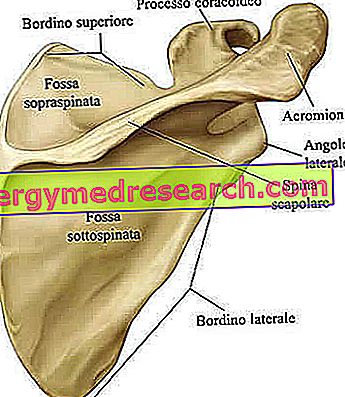
REAR SURFACE
The posterior surface of the scapula (or dorsal surface) is the bony portion turned towards the back, therefore to the opposite of the aforementioned front surface.
On the posterior surface there are at least 5 anatomical elements that deserve an accurate description: the so-called scapular spine, the supraspinatus fossa (or supraspinatus), the subpusched fossa (or infraspinata or subspina), the acromion and the crest situated under the glenoid cavity.
- Scapular spine . It is a bony prominence that crosses, mostly horizontally, the entire back surface. It cannot be said to be totally horizontal, as it tends upward in its path to the medial margin → lateral margin.
The scapular spine is important because it splits the back surface in two; the two pits that will be described in the next two points derive from this division: the supraspinatus and the sub-sinew.
- Supraspinous pit . It is the smooth area, half concave and half convex, which resides in an upper position than the scapular spine. Smaller than the subpusched fossa, it represents the point from which one of the four muscles of the rotator cuff originates: the supraspinatus (or supraspinatus) muscle.
On the medial margin it borders on the vertebral column.
- Sub-spaced pit . It is the region below the scapular spine. Larger than the supraspinous fossa, it is slightly concave in the upper and convex section in the central section. On the convex section, the rotator cuff muscle known as the sub- spinal muscle (or infraspinatus) originates.
- Acromion . It is a clear bone projection, which continues laterally (ie outward) to the scapular spine. Similar to a hook, it develops forward and has the important task of articulating with the clavicle. The articulation between the acromion of the scapula and the clavicle takes the specific name of acromioclavicular joint .
- Crest located under the glenoid cavity . Devoid of a specific anatomical name, this crest is a bone projection that extends along almost the entire lateral edge of the posterior surface, starting from the glenoid cavity. Its function is to insert a series of fibrous membranes - called fibrous or aponeurotic septa - which separate the spinal muscle from the small round and large round muscles, attached respectively to the axillary edge and to the lower angle.
As perhaps some readers will already know, the small round muscle is part of the rotator cuff muscles.

| The rotator cuff muscles and their corresponding place of origin, on the scapula. | |
| Muscle | Office: |
| Subscapularis muscle | Subscapular fossa of the anterior surface |
| In sp clear (or infraspinatus) muscle | Ditch in the back surface |
| Small round muscle (or minor teres) | Axillary (or lateral) border |
| Supraspinatus (or supraspinatus) muscle | Supraspinous pit of the posterior surface |
CORNERS OF THE SCAPOLA

The corners of the scapula are three differently pointed areas, which are located above (upper corner), laterally (lateral angle or glenoid angle) and below (lower angle).
By observing the front (or the back) surface, the reader may notice that:
- The upper corner resides in the medial position. It is covered by the trapezius muscle.
- The side angle occupies a lateral position. It takes the alternative name of glenoid angle, as it is concomitant with the glenoid fossa.
- The lower angle is medial, with respect to the glenoid angle, and lateral, with respect to the upper corner. It is covered by the large dorsal muscle and, from the dorsal side, marks the point of origin of the large round muscle.
UPPER BORDER
Visible by observing the scapula from above, the upper border runs from the upper corner to the coracoid process. Just near the coronoid process, it has a semicircular hollow, which is called the suprascapular recess .
The suprascapular nerve runs along the upper edge and then through the suprascapular cavity. The suprascapular nerve is a branching of the brachial plexus - an important reticular formation of spinal nerves - and has the task of innervating the supraspinatus muscles to the sinuous.
Of the three identifiable edges on the scapula, the upper edge is the shorter and thinner border.
Note: in addition to controlling two rotator cuff muscles, the supraspinatus nerve also has a sensory function. In fact, it is able to send sensitive information to the brain originating in the glenohumeral joint and in the acromioclavicular joint.
ASCELLAR BORDINO
The axillary border begins just below the glenoid fossa and extends obliquely, with backward orientation, to the lower corner. It is particularly important, because, on the side of the dorsal surface, it gives insertion to the initial head of the small round muscle.

Of the three identifiable edges on the scapula, the underarm border is the thickest.
MEDICAL BORDER
The medial border is the longest of the three edges of the scapula. In fact, it extends, looking at the spine, from the upper corner to the lower corner.
On the medial border, the terminal heads of 4 muscles are inserted: the anterior serratus muscle, the rhomboid large muscle, the small rhomboid muscle and the levator scapula .
Except for the first of these muscles - which is attached to the front edge by the edge - the other three all end on the side of the back surface.
OBSIFICATION OF THE SCAPOLA
At the formation of the scapula concur eight centers of ossification : one on the body, two on the coracoid process (one in a central position and one at its root), two on the acromion (one at the base and one at the outer end), one on the medial border, one on the lower corner and one on the glenoid cavity.
The ossification proceeds according to very specific moments, moments that can be summarized in these brief points:
- The process is centered on the body. This begins its activity around the eighth (8th) week of fetal life. In this phase, the future scapula looks like a quadrilateral.
The formation of an important structure (of the body), like the scapular spine, occurs around the third month of fetal life.
- At birth, the body is almost completely ossified. The glenoid cavity, the coracoid process, the acromion, the vertebral edge and the inferior angle are still cartilaginous in nature.
- Generally, between the 15th and 18th months of life, the ossification center present in the center of the coracoid process is activated. The portion to which it gives rise merges with the body around the 15th year of life.
- At about 10-11 years, the center on the glenoid cavity appears and participates in the ossification of the scapula. This center is different from the others and the doctors define it with the adjective "additional". The bone portion it gives rise to merges with the body of the scapula at the age of about 16-18 years.
- Between the 14th and 20th years of life, they enter into action, according to this temporal succession: the center at the root of the coracoid process; the center at the base of the acromion; the center on the lower corner; the center of the acromion located on the outer end; the center on the medial border.
The fusion of the various bony portions, which derive from the activity of these centers, occurs about the 25th year of life.
Functions
The scapula covers at least two fundamental functions.
The first function is to hook the upper limb to the trunk through the glenohumeral joint. The glenohumeral joint is an example of diarthrosis . Diarthroses are mobile joints, which enjoy a wide range of movement in one or more directions of space. Another important diarthrosis of the human body is the knee.
The second function of the scapula is to support the muscles involved in the movements of the shoulder joint and the muscles that end at the level of the arm.
There are 18 muscular elements that establish contact with the scapula: this shows how important the presence of the scapula is in the context of the human skeleton.
| List of the 18 muscles that originate and end at the scapula. | ||
| Muscle | Terminal end or initial end | Contact site on the scapula |
| Pectoralis minor muscle | Terminal end | Coracoid process |
| Rhomboid large muscle | Terminal end | Medial border |
| Rhomboid small muscle | Terminal end | Medial border |
| Anterior serrated muscle | Terminal end | Medial border |
| Elevating muscle of the scapula | Terminal end | Medial border |
| Trapezius muscle | Terminal end | Acromion and scapular spine |
| Subscapularis muscle | Initial end | Fossa subscapularis |
| Coracobrachial muscle | Initial end | Coracoid process |
| Long head of the triceps brachialis muscle | Initial end | Infraglenoid tubercle |
| Brief head of the biceps brachialis muscle | Initial end | Coracoid process |
| Long head of the biceps brachialis muscle | Initial end | Suprogenenous tubercle |
| Deltoid muscle | Initial end | Acromion (with median fibers) and scapular spine (with posterior fibers) |
| Supraspinatus muscle | Initial end | Supraspinous pit |
| Infraspinatus muscle (or inborn) | Initial end | Sub-spaced pit |
| Small round muscle | Initial end | Side border |
| Big round muscle | Initial end | Lower corner / side edge |
| Large dorsal muscle | Initial end | Lower angle |
| Homoioid muscle | Initial end | Top edge |
MOVEMENTS OF THE SCAPOLA
Thanks to the numerous muscles that the scapula hosts, this particular bone of the human skeleton can perform different movements, which are called:
- Elevation . It is the gesture of raising the shoulder blades.
- Depression . It is the lowering movement of the shoulder blades.
- Adduction . It is the gesture by which the two shoulder blades tend to get as close as possible to the sagittal plane.
- Abduction . It is the movement opposite to the adduction, therefore the one in which the shoulder blades tend to move as far away as possible from the sagittal plane.
- Upward rotation . It is the movement carried out by the shoulder blades when the arms are raised towards the sky.
- Downward rotation . It is the gesture carried out by the shoulder blades, when the arms are carried from above along the body.

Diseases of the Scapula
There are two relevant problems that can affect the scapula: bone fractures against it, the so-called condition known as winged shoulder blades .
FRACTURES OF THE SCAPOLA
The fracture of the scapula is a very uncommon injury.
Almost always a result of severe chest trauma, it is particularly common in people involved in car accidents and contact sports (such as soccer, rugby, ice hockey, etc.).
Usually, it does not require specific medical-surgical interventions, but only a period of absolute rest.
WING HATCHES
The clinical condition "winged shoulder blades" is when, during the pushing movements of the arm, the shoulder blades protrude backwards.

Anatomically speaking, what emerges from the scapula, when it protrudes backwards, is its medial border.