Generality
The mitral valve, or mitral valve, is located between the left atrium and the ventricle of the heart. Its job is to regulate blood flow through the orifice that connects these two cardiac compartments.
Some references to the anatomy of the heart
Before proceeding with the description of the tricuspid valve, it is useful to recall some features of the organ in which it is found: the heart .
The heart is an unequal, hollow organ made up of involuntary striated muscle tissue. Its main function is to put blood into the vessels; it is therefore comparable to a pump, which, by contracting, pushes the blood towards the various tissues and organs. It has a shape reminiscent of an inverted pyramid. At birth, the heart weighs 20-21 grams and, in adulthood, reaches 250 grams in the woman, and 300 grams in the man. The heart resides in the chest, at the level of the anterior mediastinum, rests on the diaphragm and is slightly shifted to the left. It is enveloped by the pericardium, a serofibrosis sack, which has the task of protecting it and limiting its distensibility. The heart wall is formed by three overlapping habits that from the outside to the inside take the name of:
- Epicardium . It is the outermost layer, in direct contact with the serous pericardium. It consists of a superficial layer of mesothelial cells resting on the underlying layer of dense connective tissue, rich in elastic fibers.
- Myocardium . It is the middle layer, made up of muscle fibers. Myocardial cells are called myocardiocytes. Both the contraction of the heart and the thickness of the cardiac wall depend on it. It is necessary for the myocardium to be correctly sprayed and innervated, respectively by a vasal and a nervous network.
- Endocardium . It is the lining of the cardiac cavities (atria and ventricles), consisting of endothelial cells and elastic fibers. To separate it from the myocardium, there is a thin layer of loose connective tissue.
The internal conformation of the heart can be divided into two halves: a right and a left part. Each part consists of 2 cavities, or chambers, distinct, called atria and ventricles, within which the blood flows.
Atrium and ventricle of each half are placed one above the other, respectively. On the right side, there is the right atrium and the right ventricle ; on the left side, there is the left atrium and the left ventricle . To divide neatly the atria and the ventricles of the two halves, an interatrial and an interventricular septum are present, respectively. Although the blood flow in the right heart is separated from the left one, the two sides of the heart contract in a coordinated manner: first the atria contract, then the ventricles.
The atrium and ventricle of the same half are instead in communication with each other and the orifice, through which the blood flows, is controlled by an atrioventricular valve . The function of the atrioventricular valves is to prevent the reflux of blood from the ventricle towards the atrium ensuring the unidirectionality of the blood flow. The mitral valve belongs to the left half and controls the flow of blood from the left atrium to the left ventricle. The tricuspid valve, on the other hand, lies between the atrium and the ventricle on the right side of the heart.
In the ventricular cavities, both right and left, there are two other valves, called semi-lunar valves. In the left ventricle resides the aortic valve, which regulates blood flow in the left ventricle-aorta direction; in the right ventricle takes place the pulmonary valve, which controls the flow of blood in the direction right ventricle-pulmonary artery. Like atrioventricular valves, these too must ensure unidirectional blood flow.
The tributary vessels, that is, those that carry blood to the heart, "discharge" into the atria. For the left heart, the inflowing vessels are the pulmonary veins . For the right heart, the tributaries are the superior vena cava and the inferior vena cava .
The effluent vessels, that is, those that drain blood from the heart, depart from the ventricles and are precisely those controlled by the valves described above. For the left heart, the effluent vessel is the aorta . For the right heart, the effluent is the pulmonary artery .

The blood circulation, which sees the heart as the protagonist, is the following. At the right atrium, blood rich in carbon dioxide and low in oxygen, which has just sprayed the organs and tissues of the body, arrives through the hollow veins. From the atrium, blood reaches the right ventricle and enters the pulmonary artery. Through this pathway, blood flow reaches the lungs to oxygenate and release carbon dioxide. After this operation, the oxygenated blood returns to the heart, in the left atrium, via the pulmonary veins. From the left atrium it passes to the left ventricle, where it is pushed into the aorta, that is the main artery of the human body. Once in the aorta, blood goes to flush all the organs and tissues, exchanging oxygen with carbon dioxide. Depleted of oxygen, the blood takes the venous system to return to the heart, in the right atrium, to "recharge". And so a new cycle is repeated, the same as the previous one.
The movements performed by the blood take place following a phase of relaxation followed by a phase of contraction of the myocardium, ie the heart muscle. The relaxation phase is called diastole ; the contraction phase is called systole .
- During diastole:
- The cardiac musculature of atria and ventricles, both right and left, is relaxed.
- The atrioventricular valves are open.
- The semilunar valves of the ventricles are closed
- The blood flows, through the inflowing vessels, first into the atrium and then into the ventricle. The transfer of blood does not occur in its entirety, as a portion remains in the atrium.
- During the systole:
- Cardiac muscle contraction occurs. The atriums begin, followed by the ventricles. We speak, more precisely, of atrial systole and ventricular systole:
- The amount of blood left in the atria is pushed into the ventricles.
- Atrioventricular valves close, preventing blood reflux in the atria.
- The semi-lunar valves open and the ventricular muscles contract.
- The blood is pushed into the respective effluent vessels: pulmonary veins (right heart), if it has to oxygenate itself; aorta (left heart), if it is to reach tissues and organs.
- The semi-lunar valves close after the blood has passed through them.
Diastole and systole alternate during blood circulation and the behavior of cardiac structures, regardless of whether the blood is in the right half or the left half of the heart, are the same.
To complete this overview of the heart, two other important topics remain to be mentioned. The first concerns the how and where the myocardial contraction nerve signal is born. The second concerns the vascular system that irrigates the heart.
The nervous impulse that generates the contraction of the heart is born in the heart itself. In fact, the myocardium is a particular muscle tissue, endowed with the capacity to self - control . In other words, myocardiocytes are able to generate the nervous impulse for contraction by themselves. The other striated muscles present in the human body, on the other hand, need a signal from the brain to contract. If the nerve network leading to this signal is interrupted, these muscles do not move. The heart, on the other hand, has a natural cardiac pacemaker, known as the atrial sinus node ( SA node ), at the junction of the superior vena cava and the right atrium. Generally, we talk about a pacemaker referring to artificial devices, capable of stimulating the contraction of the heart of patients suffering from certain cardiopathies. To correctly conduct the nerve impulse, born in the SA node, to the ventricles, the myocardium has other pivotal points: in succession, the signal generated by it passes through the atrioventricular node ( AV node ), for the His bundle, and for the Purkinje fibers .
The oxygenation of cardiac cells is the responsibility of the coronary arteries, right and left. They originate from the ascending aorta. Their malfunction results in ischemic heart disease. Ischemia is a pathological condition characterized by the lack or insufficient blood supply to a tissue. The blood, once the oxygen has been exchanged with the cardiac tissues, takes the venous system of the cardiac veins and coronary sinus, thus returning to the right atrium. The entire vascular network of the heart resides on the surface of the myocardium, in order to avoid their constriction at the time of cardiac muscle contraction; situation, the latter, which would alter the blood flow.
Function and anatomy of the mitral valve
The mitral valve, or mitral valve, is found in the orifice that connects the left atrium and the left ventricle of the heart. It is one of the two atrioventricular valves of the heart, together with the tricuspid one. It plays a fundamental role: it regulates the passage of blood from the atrium to the ventricle, allowing the unidirectionality of the flow at the time of the systole. During the systole, in fact, the atrium contracts, pushing all the blood into the ventricle. Only at this point, the mitral valve closes, preventing any kind of blood reflux. The diameter of the mitral valve is approximately 30 mm, while the surface of the orifice is about 4 cm2.
The opening and closing mechanism depends on the pressure gradient, ie on the pressure difference, existing between the atrial and the ventricular compartment. Indeed:
- When the blood arrives in the atrium and the atrial systole begins, the pressure in the atrium is higher than the ventricular pressure. Under these conditions, the valve is open.
- When blood arrives in the ventricle, the pressure in the ventricle is higher than in the atrium. In these conditions, the valve closes, preventing reflux.
These two situations are common to both atrioventricular valves of the heart.
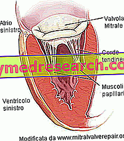
The structure of the mitral valve is composed of:
- The valve ring . Circumferential structure of connective tissue defining the valve orifice.
- Two flaps, front and back. It is said, for this reason, that the mitral valve is bicuspid . Both flaps fit into the valve ring and look towards the ventricular cavity. The anterior leaflet looks towards the aortic orifice; the posterior flap faces, instead, on the wall of the left ventricle. The flaps are composed of connective tissue, rich in elastic fibers and collagen. To facilitate the closure of the orifice, the edges of the flaps have particular anatomical structures, called commissures. There are no direct controls, of the nervous or muscular type, on the flaps. Similarly, there is no vascularization.
- The papillary muscles . They are two and are extensions of the ventricular musculature. They are sprayed by the coronary arteries and give stability to the tendinous cords.

Given the structural complexity, the proper functioning of the mitral valve depends both on the state of the flaps and the tendinous cords, and on the left ventricle. In fact, an altered morphology of the ventricle, from which the papillary muscles depart, can cause a malfunction of the mitral valve.
diseases
The most common diseases that can afflict the mitral valve are:
- Mitral stenosis. It is a narrowing of the valve orifice, caused by the fusion of the commissures or by an altered position of the tendinous cords.
- Mitral insufficiency . Incomplete closure of the valve occurs at the time of ventricular systole.
- Mitral valve prolapse syndrome, also known as mitral valve prolapse . It is an anomalous behavior of the valve leaflets, which are everted (prolapsed) towards the left altar.


