Generality
The scaphoid is the bone of the carpus which, together with the lunate (another carpal bone), forms the important articulation of the wrist.

The anatomists have coined the term scaphoid, because the bone in question has the shape of a boat.
On the scaphoid, at least 6 regions of a certain anatomical relevance are recognizable: the upper surface, which is articulated with the radium; the lower surface, which is articulated with trapezoid and trapezoid; the dorsal surface, which is home to important ligaments; the palmar surface, which houses a head of the transverse carpal ligament; the lateral surface, on which a head of the radial collateral ligament is inserted; finally, the medial surface, which is articulated with the lunate and the capitate.
Brief review of the skeletal anatomy of the wrist
The wrist is the anatomical region of the human body, which resides at the end of the distal end of the forearm and which represents the proximal portion of the hand .
The skeletal structure of the wrist is called carpus and includes 8 irregular bones, which anatomists call carpal bones or carpus bones .
The carpus bones are arranged in two rows, each containing 4 bone elements. A row is called proximal and borders on the forearm; the other row is defined as distal and confines with the 5 metacarpal bones of the hand (NB: to know the skeletal anatomy of the hand we recommend the article present here).
The bony elements of the proximal row of the carpus help to form the articulation of the wrist, thanks to their interaction with the bones of the forearm, radius and ulna ; the bony elements of the distal carpal row, instead, become part of the so - called carpometacarpal joints, thanks to their conjugation with the metacarpal bones.

What is the scaphoid?
The scaphoid is one of the 8 bones of the carpus; to be precise, it is one of the 4 bony elements of the so-called proximal carpal row.
At this point it is worthwhile to remind readers of the names of the other carpal bones and which of these bones make up the proximal row and the distal row.
In addition to the scaphoid, the other carpal bones are: the lunate, the triquatum, the pisiform, the trapezoid, the trapezoid, the capitate and the unconditioned. The lunar, triquatum and pisiform shapes are added to the scaphoid and form the proximal row of the carpus, while trapezoid, trapezoid, capitate and hooked are the elements constituting the so-called distal row (of the carpus).
In anatomy, proximal and distal are two terms with the opposite meaning.
Proximal means "closer to the center of the body" or "closer to the point of origin". Referring to the femur, for example, it indicates the portion of this bone closest to the trunk.
Distal, on the other hand, means "farther from the center of the body" or "farther from the point of origin". Referred (always to the femur), for example, it indicates the portion of this bone furthest from the trunk (and closer to the knee joint).
ORIGIN OF THE NAME
The term scaphoid derives from the Greek word scafoides (), which in Italian means "in the shape of a boat". The anatomists have chosen to use the aforementioned term, since, from the morphological point of view, the scaphoid bone resembles a small boat.
SYNONYMS OF SCAFOIDE
The scaphoid is also known as the navicular carpal bone . The use of the word "navicular bone" requires the specification "of the carpus", to avoid confusion with the navicular bone of the foot. The navicular bone of the foot is one of the 7 bones that make up the tarsus, ie the skeletal region interposed between the distal ends of the tibia and fibula (leg bones) and the 5 metatarsal bones of the foot.
Anatomy
The scaphoid is the largest bone in the proximal carpal row.
It resides on the side of the radium - which precedes it - and on the side of the thumb - which follows it.
It borders and is articulated with: radio, semi-lunar, trapezoid, trapezoid and capitate.
80% of the bony surface of the scaphoid has a covering of articular cartilage .
In describing the anatomy of the scaphoid, the experts identify in the latter at least 6 regions of a certain relevance: the upper surface, the lower surface, the dorsal surface, the palmar surface, the lateral surface and the medial surface.
UPPER SURFACE
The upper surface of the scaphoid is the region that articulates with the distal end of the radius, forming the important articulation of the wrist. It is concave, smooth and triangular in shape.
Note : there are three bony components that participate in the wrist joint: the distal end of the radius, the scaphoid (with its upper surface) and the lunate.
LOWER SURFACE
Smooth, convex and triangular, the lower surface of the scaphoid has a small ridge, which separates it into two portions: a lateral (or external) portion and a medial (or internal) portion.
The lateral portion is articulated with the trapezius, while the medial portion is articulated with the trapezoid.
In anatomy, medial and lateral are two terms of opposite meaning, which serve to indicate the distance of an anatomical element from the sagittal plane . The sagittal plane is the anteroposterior division of the human body, from which two equal and symmetrical halves are derived.
Mediale means "near" or "closer" to the sagittal plane, while lateral means "far or" farther "from the sagittal plane.
DORSAL SURFACE
The surface of the navicular on the back of the hand is called dorsal.
The dorsal surface of the scaphoid has a narrow and rough groove, which runs the entire width of the scaphoid. This groove inserts into several ligaments, including, for example, the dorsal radiocarpal ligament and the hull-lunate ligament .
PALM SURFACE
The surface of the scaphoid which is found on the palm of the hand is called palm.
Shaped, concave, the palmar surface of the scaphoid has a tubercle, which serves to hook a portion of one of the two ends constituting the transverse carpal ligament .
The transverse carpal ligament is the important band of fibrous connective tissue, which, together with the carpus bones, gives rise to the anatomical structure known as carpal tunnel
The carpal tunnel is a channel through which 9 tendons pass and the so-called median nerve ; many readers will know it, because, from its shrinkage, comes a very famous medical condition: carpal tunnel syndrome .
A few more details on the transverse carpal ligament
The transverse carpal ligament, also known as the flexor retinaculum, is a long and wide ligament, which runs along the entire wrist, from the radial side to the ulnar side. On the radial side, it finds insertion at the level of the scaphoid carpal bones and trapezius; on the ulnar side, however, it engages at the level of the pisiform and hooked carpal bones.
SIDE SURFACE
Rough and narrow, the lateral surface of the scaphoid is a region that inserts into one of the heads of the collateral radial ligament of the wrist .
MEDIUM SURFACE
The medial surface of the scaphoid is a region presenting two particular areas, which have the task of articulating with the carpal and semi-lunar bones.
Going into more detail, these two areas are called the upper articular facet and lower joint facet.
The upper articular facet interacts with the lunate; the inferior articular facet, on the other hand, relates to the capitate.
VASCULARIZATION OF SCAFOIDE
The scaphoid receives oxygenated blood from the dorsal and palmar branches of the radial artery:
- The dorsal branches of the radial artery supply about 80% of the scaphoid with blood. Most of these arterial vessels enter the bone in question at the level of that narrow and rough groove, which characterizes the dorsal surface.
The dorsal branches provide the blood supply of the entire proximal portion of the scaphoid, as well as a good part of the middle portion and of the distal portion.
- The palmar branches of the radial artery supply about 20-30% of the scaphoid blood. These arterial vessels enter the bone in question at the level of the tubercle, which characterizes the palmar surface.
The blood supply, to which the palmar branches of the radial artery supply, involves the aforementioned tubercle and the distal portion of the scaphoid.
Due to the particular distribution of blood vessels within the scaphoid, this bone is at high risk of osteonecrosis when it is fractured.
Functions
The scaphoid, together with the lunate, contributes decisively to the formation and to the movement ability of the articulation of the wrist.
Moreover, due to the position it occupies and the dimensions that characterize it, it is a fundamental element of connection between the carpal bones of the proximal row and the carpal bones of the distal row.
SUMMARY OF MOVEMENTS OF THE WRIST JOINTS
The wrist joint guarantees the hand the possibility of performing movements of:
- Bending . It is the movement that allows you to bring the palm of your hand closer to your arm. Imagining to observe an upper limb completely extended forward, the flexion of the wrist is the movement that bends the hand downwards.
- Extension . It is the movement that allows the back of the hand to be brought closer to the arm. Imagining to observe an upper limb completely extended forward, the extension of the wrist is the movement that bends the hand upwards.
- Radial deviation . It is the movement that allows the side of the hand to be brought closer with the thumb to the radio.
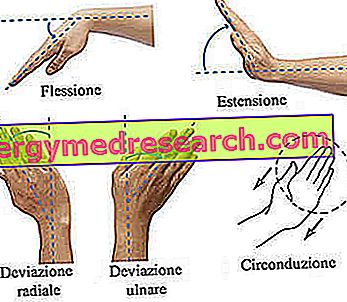
- Ulnar deviation . It is the movement that allows the side of the hand to be approached with the little finger to the ulna.
- Surrounding . It is the rotation movement of the hand.

Development
In general, the ossification process of the scaphoid occurs at the age of 6, both for men and women.
Associated pathologies
From the medical-clinical point of view, the scaphoid can be the protagonist of fractures or of injuries to the ligaments that are inserted on one of its surfaces.
FRACTURE OF SCAFOIDE
Due to the extremely critical position it occupies, the scaphoid is the carpus bone most prone to fractures.
Among the main causes of scaphoid fracture are falls with hands extended forward.
The typical symptom of a scaphoid fracture is hand pain ; if the fracture alters normal bone vascularization, it is highly probable that the scaphoid undergoes osteonecrosis.
For a certain diagnosis of fracture of the scaphoid, X-ray examination, physical examination and medical history are essential.
An inadequate treatment of scaphoid fractures - especially those characterized by osteonecrosis - favors the appearance of arthritis of the wrist.
ACCIDENTS TO SCAFOIDE LINKS
The most important injury to one of the ligaments, which are related to the scaphoid, is a medical condition known as hull-lunated instability .
Those who suffer from hull-lunated instability have a lesion affecting the hull-lunated ligament .
The hull-lunate ligament is that band of fibrous connective tissue which has the task of keeping the lunar bone of the carpus united to the scaphoid and keeping it at a constant distance.
A lesion affecting the scapho-lunated ligament involves an increase in the physiological distance that separates the scaphoid from the lunate.