Generality
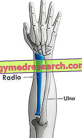
The radius is the even bone which, together with the ulna (with respect to which it is in the lateral position), forms the skeleton of each forearm.
To simplify the study, the anatomists divide it into three portions: the proximal end (or proximal epiphysis), the body (or diaphysis) and the distal end (or distal epiphysis).
The proximal end is essential to articulate with the humerus; the body accommodates numerous muscles of the forearm and of the hand; finally the distal end is relevant for its union with the proximal, scaphoid and semilunar carpal bones.
Like any other bone in the human body, radium can fracture as a result of trauma.
Definition
Radium is the even bone which, together with the ulna, constitutes the skeleton of each forearm .
The forearm is the anatomical region of the upper limb between the upper arm and the hand below.
Belonging to the category of long bones, radium forms two important joints of the human body: one with the arm bone (the humerus), called the elbow joint, and another with the carpal bones of the hand, called the wrist joint .
POSITION COMPARED TO ULNA
The radio runs parallel to the ulna, in a lateral position if the hand is facing the palm towards the observer.
Readers who do not know the concept of lateral (and its opposite meaning, ie media), are advised to consult the explanation in the box below.
Important note: meaning of medial and lateral
Medial and lateral are two terms with the opposite meaning. However, to fully understand what they mean, it is necessary to take a step back and review the concept of the sagittal plan.
Figure: the plans with which the anatomists dissect the human body. In the image, in particular, the sagittal plane is highlighted.
The sagittal plane, or median plane of symmetry, is the antero-posterior division of the body, a division from which two equal and symmetrical halves are derived: the right half and the left half. For example, from a sagittal plane of the head derive a half, which includes the right eye, the right ear, the right nasal nostril and so on, and a half, which includes the left eye, the left ear, the left nasal nostril etc.Returning to the medial-lateral concepts, the word media indicates a relationship of proximity to the sagittal plane; while the word side indicates a relationship of distance from the sagittal plane.
All anatomical organs can be medial or lateral with respect to a reference point. A couple of examples clarify this statement:
First example. If the reference point is the eye, it is lateral to the nasal nostril of the same side, but medial to the ear.
Second example. If the reference point is the second toe, this element is lateral to the first toe (toe), but medial to all the others.
IN THE LOWER ARTS IT CORRESPONDS TO ...
In the lower limb, the bone corresponding to the radius is the tibia . Together with the fibula, the tibia forms the skeleton of the leg . Like radium, tibia and fibula are two even bones.
Anatomy
Anatomy experts identify three main bone regions (or portions) in the radius: the proximal end (or proximal epiphysis), the body (or diaphysis) and the distal end (or distal epiphysis).
Anatomical meaning of proximal and distal
Proximal and distal are two terms with opposite meaning.Proximal means "closer to the center of the body" or "closer to the point of origin". Referring to the femur, for example, it indicates the portion of this bone closest to the trunk.
Distal, on the other hand, means "farther from the center of the body" or "farther from the point of origin". Referred (always to the femur), for example, it indicates the portion of this bone furthest from the trunk (and closer to the knee joint).
End? PROSSIMALE DEL RADIO
The proximal end of the radius is the bone portion closest to the arm and which, joining the latter's bone (the humerus), is the protagonist of the elbow joint.
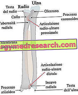
Cylindrical in shape, the proximal epiphysis has at least three relevant anatomical elements: the so-called radium head, radial neck and radial tuberosity.
- Radio head . Representing the upper apex of the radius, it is the area involved in the elbow joint. In fact, through its upper surface, it is articulated with a smooth spherical region of the humerus, which takes the specific name of capitulum .
On the medial border, it is important to point out the presence of a particular bone area, which allows the union of radius and ulna superiorly.
The head of the radio has the appearance of a disk with smooth surfaces.
- Neck of the radio . It is the bone section between the head of the radium and the radial tuberosity. Compared to the head of the radium, it has a smaller diameter; it's smooth.
- Radial tuberosity . It is a bone projection, which serves to accommodate the terminal head of the biceps brachialis muscle. Inside the forearm, it occupies a medial position, so it faces the ulna.

BODY OF THE ULNA
The body is the central portion of the radius, between the proximal end and the distal end.
Along its entire path, it has 3 surfaces - the fly surface, the dorsal surface and the lateral surface - and 3 edges - the fly, the interosseus and the dorsal.
Among the aforementioned elements, they deserve a report:
- The fly surface, as it receives the initial heads of different muscles (for example, the long flexor of the thumb and the square pronator), acts as an insertion point for the radio-carpal ligament fly and houses the so-called nutritive hole (of which we will talk later).
- The dorsal surface, because it gives rise to the important thumb muscles.
- The lateral surface, as it houses the initial ends of the supinator and pronator round muscles of the forearm.
- The interosseous border (or interosseous crest ), because it engages the so-called interosseous radio-ulnar membrane . The radio-ulnar interosseous membrane is a thin sheet of fibrous tissue, interposed between radius (medially) and ulna (laterally), which indirectly unites the two bones of the forearm.
Bone unions involving interosseous membranes are a particular type of fibrous joints, identified with the name of syndesmosis .
Unlike the body of the ulna - which tends to tighten in a distal direction - the body of the radium tends to widen.
Syndesmosis
All the fibrous joints, which hold the bone elements together, are part of the syndesmosis category, an interosseous membrane (like the one mentioned above) or a network of ligaments.
Another important fibrous articulation, very similar to that present between radium and ulna, is the syndesmosis located between tibia and fibula.
End? DISTALE DEL RADIO
Wide and quadrangular, the distal end of the radius is the bone portion closest to the wrist and with which it takes part in the formation of the homonymous articulation.
Its most important anatomical components are:
- The styloid process, in a lateral position;
- The ulnar groove, on the medial surface;
- The two facet joints, on the surface in contact with the bones of the carpus.
The styloid process is an evident conical bone projection, on which they find insertion: the terminal head of the brachioradial muscle (NB: originates in the humerus), the initial ends of some thumb muscles (therefore of the hand) and one of the two Heads of the radial collateral ligament of the wrist.
The ulnar cavity is a depression (that is, a concavity), in which it perfectly houses the lateral surface of the ulna's head. This radio-ulna contact in the distal area is added to the radio-ulna union in the proximal area, described above.
Finally, the two facet joints - one lateral and one medial - are the junction points with two of the 8 carpal bones of the hand, the scaphoid and the lunate . In this case, the lateral surface is articulated with the scaphoid, while the medial surface with the lunate.

BLOOD SPRAYING
Internally, long bones, such as radium (but also the ulna or humerus), have a very specific network of arteries and veins, which serves to guarantee them the right supply of oxygen and nutrients.
Arteries - that is, in vessels that carry oxygen-rich blood - are the so-called nutritive artery and periosteum arteries ; the veins - that is, the vessels that drain the oxygen-poor blood - are the so-called nutritive vein and the periosteum veins .
In the case of radium, the aforementioned arteries derive from the anterior interosseous artery, while the aforesaid veins flow into the anterior interosseous vein .
The nutritive artery and the nutritive vein deserve a particular note, as they penetrate the body of the radium, through a previously named structure: the nutritive hole (also known as nutritious channel).

Figure: The nutritious vessels and the nourishing hole in the long bones.
RADIO ARTICULATION
In summary, there are four ways in which the radio participates:
- The elbow joint, fruit of the union between the head of the radium and the capitulum of the humerus.
- The upper radio-ulnar joint, which joins the radium head, located on the proximal end of the radius, to the ulnar cavity, located on the proximal epiphysis of the ulna.
- The inferior radio-ulnar joint, which joins the distal end of the radium (in this case, the ulnar groove of the radius) to the distal end of the ulna (to be precise, the head of the ulna).
- The articulation of the wrist, result of the collaboration between the two articular facets of the ulna and the carpal, scaphoid and semilunar bones.
OBSIFICATION OF THE RADIO
Four ossification centers contribute to the formation of radium: one centered on the body, one on the proximal epiphysis, one on the distal epiphysis and finally one on the radial tuberosity.
To begin the process of ossification is the center present on the body of the radium; to follow and in succession, the center of the distal end, the center of the proximal end and the center of the radial tuberosity come into action.
Going into more detail:
- The ossification center of the body is activated around the 8th week of fetal life. Its activity causes the bone to develop towards the body and the ends.
- The ossification center of the distal end is set in motion between the eighth and 26th month of life. The bone portion it gives rise to meets the bony portion of the ossification center of the body at about the 20th year of life.
- The ossification center of the proximal end begins its activity at about the 5th year of life. The resulting bone portion meets the bony portion of the body around the 17th-18th year of life.
- The ossification center of the radial tuberosity (NB: is an additional center) appears and comes into action between the 14th and 15th year of life.
Functions
Radium is a fundamental element in the formation of the elbow and wrist joints. Moreover, it welcomes those muscles of the forearm and of the hand that allow to fully exploit the potential of the aforementioned articular elements.
Together with the humerus (and its joints), the ulna and the skeleton of the hand, the radius makes it possible to lift weights, hold something, write, throw objects, etc. Therefore, it is extremely important in many everyday activities.
| List of muscles that originate and end at the radius. | ||
Muscle | Head end or initial leader | Contact site on the tibia |
| Biceps brachialis muscle | Head end | Radial tuberosity |
| Supinator muscle | Head end | Lateral surface of the body of the radium |
| Pronator square muscle | Head end | Last front section of the radio body |
| Brachioradial muscle | Head end | Styloid process |
| Long abductor muscle of the thumb | Initial leader | Dorsal surface of the body of the radium |
| Short extensor muscle of the thumb | Initial leader | Dorsal surface of the body of the radium |
| Superficial flexor muscle of the fingers | Initial leader | Fly surface of the radio body |
| Long flexor muscle of the thumb | Initial leader | Fly surface of the radio body |
| Pronator round muscle | Initial leader | Lateral surface of the body of the radium |
Diseases of the Radio
Like practically any other bone in the body, the radium can fracture as a result of traumas against it.
There are various types of radium fractures: the Colles fracture, the radius head fracture, the Smith fracture, the Barton fracture, the Monteggia fracture and the Galeazzi fracture.
* Please note: the proper names belong to the doctors who described this type of fracture for the first time.
COLLES FRACTURE
The Colles fracture concerns the distal end of the radius and involves the dislocation of the wrist joint on the dorsal side of the upper limb.
In most cases, the cause is a fall with the extended hand.
FRACTURE OF RADIO HEAD
The fractures of the radium head derive from impacts such that the head of the radium collides with the capitulum of the humerus, breaking.
Generally, the cause is a fall with the extended hand.
SMITH FRACTURE
Smith's fracture is the rupture of the distal end of the radium from the palm side of the hand.
This results in the dislocation of the wrist joint on the front side of the upper limb (so the effects are contrary to those of the Colles fracture).
Generally, the cause is a fall on the back of the hand.
BARTON FRACTURE
Barton's fracture is an intra-articular rupture that affects the distal end of the radius. The consequences are the inferior radio-ulnar joint.
MONTEGGIA FRACTURE AND GALEAZZI FRACTURE
The fracture of Monteggia and the fracture of Galeazzi have in common the fact that, often, they involve both bones of the forearm, therefore both radio and ulna.
To cause this double fracture are traumas whose impact force is transmitted from bone to bone, through the interosseous membrane.
The Monteggia fracture is a clinical condition characterized by the rupture of the ulna's body and a dislocated radium head.
Galeazzi's fracture, on the other hand, is a fracture of the distal end of the radius, combined with a dislocation of the ulna's head (NB: unlike radium, the head of the ulna takes place on the distal epiphysis).