Generality
The crural hernia, or femoral hernia, is a type of abdominal hernia, characterized by the protrusion of a bowel of the abdomen in the so-called femoral canal. The femoral canal is a short longitudinal duct, located approximately at the level of the groin, near the femoral artery and the femoral vein.

The factors that can induce a collapse of the muscular wall of the abdomen are different.
If mild, crural hernia is often asymptomatic; if, on the other hand, it is severe, it can cause various problems (swelling, groin pain, etc.), some of which are also very serious and life-threatening.
In case of severe crural hernia, an early diagnosis and a timely ad hoc surgical procedure are necessary.
Mild cases of crural hernia generally do not require any type of treatment.
What is a hernia
A hernia is the outflow of a bowel and / or adjacent tissues (for example the surrounding fatty tissues) from the body cavity that normally contains these anatomical elements (NB: the word viscera indicates a generic internal organ).
The spill may be total or partial.
What is the crural hernia
The crural hernia, or femoral hernia, is a type of abdominal hernia, in which a bowel of the abdomen has escaped from its cavity and has penetrated into the femoral canal .
Described as the medial compartment and smaller than the so-called femoral sheath, the femoral canal resides in the highest and innermost part of the leg, near the groin.
Because of its particular location, the crural hernia is very reminiscent of the inguinal hernia; however, it is worth pointing out immediately that the two conditions are quite distinct and the episodes of crural hernia are more at risk of complications.
SOME ADDITIONAL INFORMATION ON THE FEMALE CHANNEL
About 1.3 cm long (therefore very short) and in a longitudinal position with respect to the body, the femoral canal borders on:
- The inguinal ligament, anteriorly
- The pectine ligament, posteriorly
- The lacunar ligament, medially
- The femoral vein, laterally
Its upper mouth is called the femoral ring ; to close the femoral ring is a band of connective tissue, called the femoral septum . Through the femoral septum, the lymphatic vessels that run along the femoral canal pass; above the septum is the intestinal compartment.
Within the femoral canal, there are:
- Deep inguinal lymph nodes
- The lymphatic vessels that drain the lymph that reaches the deep inguinal lymph nodes
- Empty space
- Loose connective tissue
The empty space in the femoral canal allows the nearby femoral vein to relax more easily, while carrying blood from the periphery to the heart.
The femoral sheath - which includes the femoral canal - forms, along with the femoral nerve, the great saphenous vein and the sartorius muscles, a long adductor, ileopsoas and pectineus, an anatomical region known as the femoral triangle (Scarpa's otriangolo).
Size of the femoral canal
- Length: 1.25-1.3 cm
- Width (at the level of the femoral ring): 1.25 cm
Epidemiology
Crural hernia mainly affects women. The reason for this is probably to be found in the particular shape of the female pelvis, which seems to predispose to the problem.
Affected subjects generally have 30-40 years of age or older.
The appearance of a crural hernia in children is a very uncommon event.
Causes
Abdominal hernias arise due to a collapse of the muscular wall of the abdomen ; this wall serves and contain the abdominal organs in their original location.
In the specific case of a crural hernia, the failure involves the muscular region of the abdomen which is near the femoral canal, just above the femoral ring.
WHAT HAPPENS AFTER THE SALE OF THE ABDOMINAL WALL
With the collapse of the abdominal wall overlying the femoral canal, the viscera (generally a tract of intestine), which resides there above, tends to slip into the crack created by the same yielding. After passing through the fissure, cross the femoral ring and wedge into the underlying conduit.
WHAT CAN THE DETERMINATION OF THE ABDOMINAL WALL DETERMINE?
To favor the collapse of the abdominal wall above the femoral canal it can be:
- The presence of a particularly weak abdominal musculature . This condition is the main cause of crural hernia in infants and young children, who still have weak abdominal muscles. With the physiological strengthening of these muscles during growth, femoral hernias tend to disappear naturally.
That said, it should be pointed out that even adult people may have abnormally weak abdomen-prone musculature.
- Repeated lifting of very heavy objects .
- Excessive strains in the toilet due mainly to the presence of constipation
- Severe obesity
- Strong and repeated coughing
- Enlarged prostate . This is an exclusive factor favoring the male sex.
- The state of pregnancy . This is an exclusive factor favoring the female sex.
Symptoms and Complications
The less severe forms of crural hernia are often asymptomatic . In other words, they lack symptoms and signs that can, in some way, reconnect to a problem in the area that corresponds to the femoral canal.
Quite the contrary, the moderate-severe forms of crural hernia induce the appearance of a precise symptomatology, which includes:
- The presence of a bulge near the groin . This swelling is palpable and, in the most severe cases, also easily visible.
Generally, it tends to disappear, temporarily, if pressed or if the patient assumes a horizontal position; while it tends to be accentuated, always temporarily, by coughing or during physical exertion.
- Pain in the groin
- Painful sensation that intensifies when the patient stands up, lifts heavy objects or performs an intense physical effort.
- Hip pain . This disorder appears only when the crural hernia is particularly close to the joint linking the acetabulum to the femoral head.
COMPLICATIONS
A crural hernia can become a life-threatening condition in two circumstances:
- When it comes out of the abdominal wall is a portion of the intestine and this portion suffers an occlusion ( intestinal obstruction ). An intestinal obstruction prevents the bowel contents from advancing normally and this is the cause of some characteristic symptoms, such as: nausea, vomiting, pain (or cramping) in the stomach.
- When the herniated (that is, escaped) bowel suffers a " constriction ". With the term "bottleneck", doctors identify the situation in which the herniated tract of the bowel no longer receives the correct blood supply. Without the right blood supply, the cells of the portion involved in the spillage undergo death (or necrosis), lack of oxygen and nourishment.
The "choking" of a crural hernia represents a medical emergency to be treated with extreme timeliness.
Its most typical symptom is the strong and sudden pain that goes from the lower abdomen to the groin area affected by the hernia.
In general, mild and still persistent crural hernias give rise to complications only in rare cases.
Conversely, moderate to severe forms of crural hernia are at high risk of complications.
Diagnosis
In most cases, an accurate physical examination is sufficient to identify the presence of a crural hernia. By physical examination we mean the evaluation by the doctor of the symptoms and signs that the patient reports.
If the doctor has doubts or wants, for a given reason, to deepen what is observed with the physical examination, he can resort to an ultrasound of the abdominal and inguinal areas .
When the crural hernia is asymptomatic, its identification is often random and occurs during medical checks performed for other reasons.
EXAMINATION OBJECTIVE
A thorough physical examination requires the doctor to visit the patient and ask him about the symptoms.
The most common questions - because they are more important for a correct diagnostic analysis - are:
- What are the symptoms?
- When did the first clinical manifestations appear?
- Are there moments or particular circumstances that increase or reduce the symptoms?
- Are there any particular signs at the level of the painful area?
At the end of such a questionnaire, the doctor analyzes the painful area in first person, palpating it and assessing whether there are any significant swelling or other anomalies.
ULTRASOUND
An ultrasound is a non-invasive diagnostic imaging procedure, which allows, by means of an ultrasound probe, to observe the internal parts of the body.
In the presence of a suspected crural hernia, the doctor performs the ultrasound examination in the region between the abdomen and groin, to assess what is the appearance of the internal tissues (in particular the muscular tissues) and of the underlying organs.
If there is actually a crural hernia, the probe shows, on an appropriately connected monitor, the collapse on the muscular wall and the viscus protruding into the femoral canal.
Treatment
Asymptomatic and small crural hernia does not require special treatment.
In fact, in these situations, the doctors simply recommend a periodic monitoring of the visceral leak, as a precautionary / preventive measure.
The situation changes radically in the presence of symptomatic, large and / or induced crural hernias. In fact, in such circumstances, doctors must resort to ad hoc surgery, which involves replacing the herniated (that is, leaked) bowel in its original location and sealing the fissure formed on the abdominal wall.
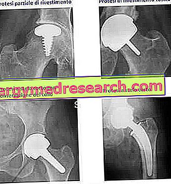
There are two possible surgical approaches: traditional (or " open ") and laparoscopic .
Surgery for the resolution of a crural hernia can last from a minimum of 30 to a maximum of 45 minutes.
GENERAL INSTRUCTIONS FOR INTERVENTION
Regardless of the approach used, crural hernia surgery generally involves a series of pre-operative clinical tests, a questionnaire related to the patient's clinical history, the suspension of certain pharmacological treatments (if any) and anesthesia general.
Going further into details:
- Usually carried out a week before the procedure, pre-operative clinical tests consist of: blood tests, electrocardiograms, blood pressure measurements and urinalysis.
If the outcome of these assessments is positive, it means that there are the conditions to intervene on the patient.
- The clinical history questionnaire allows the doctor to know if the patient suffers from some chronic disease or is allergic to certain drugs or anesthetics.
- The suspension of any pharmacological treatment based on antiplatelet agents (aspirin), anticoagulants (warfarin) and anti-inflammatory drugs (NSAIDs) serves to reduce the risk of bleeding. The aforementioned drugs, in fact, reduce the coagulation capacity of the blood and this predisposes to large bleeding.
- General anesthesia serves to render the patient unconscious, therefore insensitive to pain.
It has an important implication: on the day of the intervention, the patient must appear in complete fasting for at least 6-8 hours. Thus, the last meal allowed is that of the previous evening.
Within this framework, water is an exception, whose intake is granted up to a couple of hours before the operation.
TRADITIONAL OR "OPEN HEIGHT" SURGERY INTERVENTION
The traditional surgical procedure is a somewhat invasive operative method, which involves a single incision of 3-4 centimeters on the lower abdomen, on the side where the crural hernia resides.
Through this incision, the surgeon acts directly on the outgoing viscus and closes the fissure generated on the abdominal wall.
For closure (or sealing or patching), use a metal mesh, which applies to the failure area.
At the end of the sealing, it closes the starting incision with absorbable sutures.
In the absence of obstructions or bottlenecks, the patient can return home the same day as the surgery, after a few hours of precautionary hospitalization.
LAPAROSCOPIC SURGERY INTERVENTION
Surgery with a laparoscopic approach is a minimally invasive operative method, which involves the practice of three mini incisions on the abdomen: one of 1.5 cm and two of 0.5 cm.
Through the larger opening, the surgeon inserts a special instrument, called a laparoscope, which is essential for the correct execution of the procedure.
Through the smaller openings, it inserts the surgical instruments for the replacement of the herniated viscera and for the sealing of the abdominal wall sold.
After completing the most important part of the operation, it closes the small incisions with a few resorbable stitches.
As in the case of "open" surgery, in the absence of occlusion or bottlenecks, the patient can return home at the end of a precautionary hospitalization lasting a few hours.
The laparoscope: what is it?
The laparoscope is the main and most representative instrument of laparoscopy (or laparoscopic surgical technique).
Similar to a drinking straw, at the end to be inserted in the abdomen, it presents a light source and a camera connected to an external monitor.
Together, these two devices allow the operating surgeon to illuminate the abdominal cavity and to orient itself inside it, while using the other surgical instruments.
THE TWO APPROACHES TO COMPARE
Recently, after several studies, the doctors agreed that the two operational approaches are equivalent, in terms of advantages and disadvantages.
In fact, compared to "open" surgery, the laparoscopic procedure has both points in favor and points against it.
Starting from the advantages, these consist of:
- Minimal surgical incisions and very rapid wound healing.
- Very low blood loss and very low risk of severe bleeding.
- Short recovery times.
Moving on to the disadvantages, these are:
- The greatest risk of injury to internal organs, through surgical instrumentation.
During "open-air" operations, this risk is lower because the surgeon has a better view of the internal anatomy of the abdomen and of the movements that he must perform to repair the hernia, without damaging the tissues and the neighboring organs.
- The highest cost of the intervention. The instrumentation used (laparoscope, etc.) is more complex and expensive, therefore it is not for everyone.
As regards the danger complications, the two operating methods are similar in this respect too.
What affects the choice of the intervention method?
The factors that influence the choice of the method of intervention are substantially two: the state of health of the patient - which if he is healthy and not old can "hold" the general anesthesia (therefore the laparoscopy) - and the experience of the surgeon in a certain operational method.
IN THE EVENT OF OCCLUSION OR THINNING
In the presence of an intestinal obstruction and / or a "narrowing" that has irreparably damaged the intestine, the surgeon must intervene in an invasive manner and eliminate the intestinal region which is no longer functional and incurable .
If for any reason this does not happen, the patient would be in danger of life.
After a crural hernia operation that forced the elimination of part of the intestine, hospitalization can last up to 4-5 days, confirming how delicate the aforementioned situation is.
THE FIRST POST-OPERATIVE FEELING
Once the most important effects of anesthesia have disappeared, it is likely that the patient will experience pain at the operated area. This is a completely normal sensation, which can last a few days and for which doctors advise taking paracetamol (or a painkiller ).
IMPORTANCE OF PERSONAL HYGIENE
Doctors recommend maximum personal hygiene in order to reduce the risk of infection.
Precisely for this reason, just before discharge, a qualified member of staff explains to the patient all the rules for washing without wetting the dressings and to keep wounds clean.
RECOVERY TIMES
The return to normal daily activities must be gradual and must take place according to the sensations felt; in other words, it is good not to force the recovery and, if you feel pain while you are carrying out a certain effort, stop immediately.
In general, for the resumption of lighter daily activities, it is sufficient to wait 1-2 weeks, while for the resumption of heavier activities, it is necessary to wait 4 to 6 weeks.
The return to work depends on the work activity itself: if the patient performs a sedentary job, 1-2 weeks of rest are enough; if, instead, the patient carries out manual work, it takes several more weeks, sometimes even 6.
To resume driving, it is advisable to wait until sitting at the wheel does not create any pain or discomfort.
Prognosis
The prognosis depends on the severity of the crural hernia.
A mild crural hernia is generally without consequences and very rarely leads to complications.
On the contrary, a severe crural hernia can be very annoying and has a good chance of giving rise to complications.
RISKS OF RECIDIVES
According to some statistics, 1-5% of people suffering from crural hernia develop a recurrence (ie they suffer from a crural hernia again).