Generality
Osgood Schlatter's disease is knee pain, which arises from inflammation of the tibial tuberosity.

Also known as tibial tubercle apophysitis, Osgood Schlatter disease is a purely juvenile medical condition, with the occurrence of bone immaturity of the tibial tuberosity, typical of the adolescent years, and an abnormal mechanism of traction of the patellar tendon, in the comparisons of the tibial tuberosity itself.
The typical symptoms of Osgood Schlatter disease are pain and swelling just below the knee, to be precise where the patellar tendon engages the tibial tuberosity.
In general, the diagnosis of Osgood Schlatter disease is based on physical examination and medical history.
In almost all cases, Osgood Schlatter disease is a temporary condition, which resolves spontaneously at the end of bone development typical of adolescence.
What is the Osgood Disease Schlatter?
Osgood Schlatter disease is the medical condition characterized by knee pain, which arises from an inflammatory suffering in the tibial tuberosity, ie the anterior prominence of the distal epiphysis of the tibia on which the patellar tendon is inserted.
Also known as tibial tubercle apophysitis, Osgood Schlatter disease is a purely juvenile disorder: the conditions for suffering tibial tuberosity, in fact, exist only in the years of greater skeletal growth.
Experts do not consider Osgood Schlatter's disease a serious condition, as it is destined to resolve spontaneously within a few months.
Osgood Schlatter disease is an example of apophysitis ; in medicine, the term "apophysite" refers to a variety of osteochondrosis, characterized by the inflammation of an apophysis, that is, a projection or an outgrowth of a bone of the human body.
To understand: brief review of Tibial Tuberosity and Patellar Tendon
TIBIAL TUBEROSITY
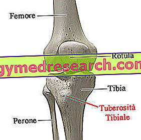
The tibial tuberosity (or anterior tibial apophysis ) is the bony prominence detectable by touch, which emerges on the front face of the proximal epiphysis of the tibia (leg bone), just below the so-called condyles, ie the enlarged areas that constitute the apex of the bone in question.
In anatomy, the term " leg " identifies the part of the lower limb that runs from the knee to the ankle.
The skeleton of the leg includes two bones: the tibia and the fibula .
ROTULEO TENDON
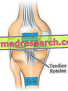
The patellar tendon, or patellar ligament, is the band of fibrous connective tissue, which joins the patella (knee) to the characteristic bony prominence, called tibial tuberosity, present on the front face of the proximal epiphysis of the tibia (upper part of the leg, just below the knee).
Structural component of the knee joint, the patellar tendon has the peculiarity of being in continuity with the tendon complex, which joins the quadriceps femoral muscle to the patella.
Located on the anterior surface of the thigh, the quadriceps femoris muscle is a muscular formation that includes as many as 4 elements, which are: the vast lateral muscle, the intermediate broad muscle, the vast medial muscle and the rectus femoral muscle.
Flat, broad and about 4.5 centimeters long, the patellar tendon has the task of keeping the patella in the correct position and of supporting the quadriceps femoris in the action of extending the knee.
Did you know that ...
The patellar tendon is a source of connective tissue useful for the surgical repair of ligaments that, once damaged, lack the ability to heal spontaneously (eg, anterior cruciate ligament of the knee).
Epidemiology
Osgood Schlatter disease is the leading cause of knee pain in young people.
Reliable statistical studies say that Osgood's disease Schlatter:
- At some point in life, it affects 4% of the general population residing in the Western world;
- It affects young people especially during the years of greater bone growth (therefore under 16, in males, and under 14, in females);
- It is more common in young males than in young females; in particular, males between the ages of 10 and 15 suffer the most.
Origin of the name
Osgood Schlatter's disease owes its name to Robert Bayley Osgood (1873-1956), an American orthopedic surgeon, and Carl B. Schlatter (1864-1934), a Swiss surgeon, who deserve credit for having described first, independently of each other, the condition in question.
Causes
Premise…
During the bending and extension movements of the knee, performed in a normal walk or a run, the quadriceps femoral muscle of the thigh is the protagonist of a contraction that affects the patellar tendon and indirectly on the tibial tuberosity; the patellar tendon, in fact, is subjected to a tension such that it exerts traction against the prominence that unites it to the tibia.
What happens in Osgood's Schlatter Disease?
Concurrent with the onset of Osgood Schlatter disease are the bone immaturity of the tibial tuberosity, typical of the adolescent years, and an abnormal mechanism of traction of the patellar tendon towards the tibial tuberosity itself. In subjects with Osgood Schlatter disease, in fact, the condition stems from an unusual stress, on the part of the patellar tendon, of a tibial tuberosity not yet completely ossified, such as that present in adolescents of 10-15 years.
The consequences
In young people with Osgood Schlatter disease, the abnormal behavior of the patellar tendon inflames the tibial tuberosity which is not yet completely ossified and induces its displacement from its natural site to an alternative site.
The migration of the tibial tuberosity involves a change in the area of ossification of the latter as well as the formation of an abnormal protuberance below the knee (anomalous because it is absent in people spared from Osgood's disease Schlatter).
As will be seen later, in the presence of a correct therapeutic management of the condition, the aforementioned anomalous protuberance and the displacement of the ossification site of the tibial tuberosity have no future repercussions on the functionality of the patellar tendon, knee and / or quadriceps femoral muscle.
Risk Factors: What Does Osgood Disease Help Schlatter?

Experts have observed that, to favor Osgood Schlatter's disease, they are:
- The practice of sports where racing and jumps are prevalent;
- A certain genetic predisposition to the development of the condition in question;
- An imbalance between skeletal growth and the growth of the muscle-ligament apparatus, such that bone growth anticipates, as it is faster, the growth of muscles and ligaments.
Symptoms and Complications
The characteristic symptoms of Osgood Schlatter disease are pain and swelling just below the knee, where the patellar tendon engages the tibial tuberosity.
Characteristics of Pain
Osgood Schlatter disease produces pain that worsens with physical activity and improves with rest .
It should be noted that the aforementioned pain increases in intensity even when the tibial tuberosity involved is subject to trauma; this is a confirmation of the suffering of the tibial tuberosity itself.
Osgood Schlatter disease: Mono- or Bilateral condition?
Osgood Schlatter disease can be either a unilateral condition - that is, it produces symptoms on only one lower limb - or a bilateral condition - that is, it induces symptoms on both lower limbs.
According to some statistical studies, Osgood Schlatter disease causes bilateral symptoms in 20-30% of patients.
Complications
In the most serious cases, Osgood Schlatter disease can lead to a fracture of the distal tibia of the tibia following very intense stresses from the tibial tuberosity.
It is important to point out that this complication is very rare.
Diagnosis
In general, the diagnosis of Osgood Schlatter disease is based on the analysis of symptoms and signs ( physical examination ) and on clinical history (or medical history ).
Sometimes, however, it happens that doctors prescribe a knee x- ray ( knee X-rays), with the aim of analyzing in detail the consequences of the condition on the tibial tuberosity.
How to predict Osgood Schlatter disease: Knee echography
Doctors specializing in the diagnosis and treatment of musculoskeletal disorders in young people have found that knee ultrasound is an instrumental test capable of detecting Osgood Schlatter disease at the beginning, when the symptoms are still not present. In fact, knee ultrasonography can detect, on the cartilage that constitutes the tibial tuberosity, a typical anomaly produced by Osgood Schlatter's disease during onset.
Differential diagnosis
In the presence of Osgood Schlatter disease, the differential diagnosis involves the distinction of the condition just mentioned by a syndrome, known as Sinding-Larsen and Johansson syndrome, which involves the patellar tendon where it attaches to the patella.
Therapy
In almost all cases, Osgood Schlatter disease is a temporary condition, which resolves spontaneously at the end of the peak of bone growth typical of adolescent age (16 years, in males, and 14 years, in females).
Waiting for this to happen, doctors recommend a series of conservative remedies, including:
- Rest from activities that produce pain. If the patient suffers particularly during activities that involve running or jumping, it is good to temporarily address him to physical practices that do not cause pain;

- Apply ice to the painful area for 15-20 minutes, repeated at least 4-5 times a day. Ice packs possess an important anti-inflammatory power, which, however, many underestimate;
- Stretching and muscle strengthening exercises, aimed at improving the elasticity and tone of the quadriceps femoral muscle. Experts have observed that a more elastic and stronger quadriceps femoral muscle reduces the effects that the abnormal tension of the patellar tendon produces against the tibial tuberosity;
- The assumption, at times of greatest pain, of an anti-inflammatory with pain-relieving properties. Among the indicated anti-inflammatories, include paracetamol and ibuprofen (an NSAID);
- Use of a knee brace with support function for the patellar tendon.
The doctors observed that putting into practice the aforementioned conservative remedies avoids complications (fracture of the tibia) and ensures a resolution without the consequences of the condition; on the other hand, neglecting the aforementioned favors the risk of fracture of the tibia and increases the chances of having to resort to surgery for remodeling of bone anomalies, formed after the displacement of the tibial tuberosity.
Schlatter's disease tends to modify the anatomy of the tibial tuberosity, but, with the right conservative remedies, this does not involve any future dysfunction of the knee, the patellar tendon and / or the quadriceps femoral muscle.
What to do if the symptoms persist?
If the symptoms of Osgood Schlatter disease persist even at the end of the adolescent peak bone growth, it is very likely that the patient will have to undergo surgery .
In short, surgical treatment of Osgood Schlatter disease involves the removal of osseous abnormalities, formed by ossification, following the displacement of the tibial tuberosity.
This therapy is not particularly complex and guarantees optimal results; after a few weeks of rest and physiotherapy, in fact, the patient can return to perform any type of physical activity.
The use of surgery to resolve Osgood Schlatter's disease is very rare.
Prognosis
The prognosis in the case of Osgood Schlatter disease is generally benign; only in rare cases, in fact, the condition can give rise to complications or make it necessary to resort to surgery.
If the patient adheres to the indications of the doctors regarding rest, ice application etc., Osgood Schlatter's disease tends to have no long-term repercussions.