Generality
The electrocardiogram, or ECG, is a diagnostic test, which involves the use of an instrument capable of recording and graphically reporting the rhythm and electrical activity of the heart.
The electrocardiogram instrument is the electrocardiograph.

Moreover, it allows to evaluate the functioning of a pacemaker or an implantable cardioverter defibrillator, in all those subjects who are carriers of devices for the normalization of the cardiac rhythm.
There are three types of electrocardiograms: the ECG at rest, the dynamic ECG according to Holter and the exercise ECG.
Cardiologists are able to understand what is the state of health of the heart and its functioning by the appearance of the electrocardiographic tracing.
What is the electrocardiogram?
The electrocardiogram, or ECG, is an instrumental diagnostic test that records and reports the rhythm and electrical activity of the heart.
The instrument used for the electrocardiogram is called an electrocardiograph .
The doctor who is usually responsible for the interpretation of an electrocardiographic trace is a cardiologist, that is a doctor specialized in cardiology.
BRIEF ANATOMICAL AND FUNCTIONAL CALL OF THE HEART
The heart is an unequal organ, which can be divided into four cavities (the right atrium, the left atrium, the right ventricle and the left ventricle) and composed of a very particular muscle tissue: the myocardium .
The peculiarity of the myocardium lies in the ability to generate and conduct nerve impulses by the contraction of the atria and the ventricles.
The source of these impulses, which are comparable to electrical signals, resides at the level of the right atrium and is called the atrial sinus node .
The atrial sinus node has the task of scanning the right frequency of contraction of the cardiac organ (the so-called heart rate ), in such a way as to guarantee a normal heart rhythm.
The normal heart rhythm is also called sinus rhythm .
uses
The electrocardiogram allows the cardiologist to detect:
- The presence of cardiac arrhythmias .
A cardiac arrhythmia is an alteration of the normal heart rhythm (sinus rhythm).
The normal heart rhythm of an adult human being has a resting contraction frequency of between 60 and 100 beats per minute.
- A myocardial ischemia or infarction, probably secondary to a narrowing or complete occlusion of the coronary arteries of the heart (NB: myocardial infarction and heart attack are synonymous).
The coronary arteries of the heart are the arterial vessels that supply the myocardium with oxygenated blood and nutrients.
In medicine, the narrowing and complete occlusion of the coronary arteries of the heart take the generic name of coronary artery disease or coronary disease.
- The presence of structural alterations of the cardiac cavities, atria and / or ventricles .
The structural alterations of the cardiac cavities include conditions such as: dilated cardiomyopathy, hypertrophic cardiomyopathy, left ventricular hypertrophy and enlarged heart.
In such circumstances, the walls of atria and / or ventricles can thicken or stretch.
- The outcomes of a previous heart attack .
The heart attack leaves indelible signs both anatomically and functionally.
Patients who have suffered a myocardial infarction must undergo periodically an electrocardiogram, to monitor the condition of their heart.
- The presence of cardiac conditions, characterized by an alteration of the electric conduction . Some examples of these cardiac conditions are: long QT syndrome and branch blocks (right or left).
Furthermore, the electrocardiogram allows to evaluate:
- The functioning of pacemakers and similar devices (such as the implantable cardioverter defibrillator ), in subjects who are clearly carriers of it.
- The effects on the heart of those drugs that could alter, in some circumstances, the frequency or electrical conduction of the heart.
Preparation
In general, the electrocardiogram does not require any special preparation.
However, it should be noted that patients undergoing pharmacological treatments or carriers of pacemakers (or similar instruments) must communicate this condition to their cardiologist.
Procedure
There are three main types of electrocardiogram:
- The electrocardiogram at rest (or basic electrocardiogram)
- The electrocardiogram according to Holter (or dynamic electrocardiogram according to Holter)
- Effort electrocardiogram
Before analyzing each type of electrocardiogram, it is good to clarify what an electrocardiograph is.
An electrocardiograph is a computerized device that, through a series of electrodes, records the cardiac function and translates it graphically onto a monitor or a sheet of graph paper.
The graph obtained after recording the cardiac function is called a path .
In a generic path, lines describe the rhythm and electrical activity of the heart, which in the medical jargon take on the word " waves ".
The appearance of the waves and the distance between them are the elements of the path that allow cardiologists to interpret the state of health of the heart under examination.
The last fundamental information on the electrocardiograph concerns the time and speed of the apparatus in drawing the path. The speed with which the electrocardiograph proceeds to bring the waves back onto the sheet of graph paper is 25 millimeters per second (25 mm / sec).
In light of this, it must be taken into account that:
- Each square of 1 millimeter of the graph paper corresponds to 0.04 seconds.
- 5 squares of 1 millimeter each (in all 5 millimeters) correspond to 0.2 seconds (0.04 * 5 = 0.2).
- 5 large squares correspond to 1 second (0.2 * 5 = 1).
RESTING ELECTROCARDIOGRAM
Before the resting electrocardiogram begins, a doctor's assistant - usually a nurse - invites the patient to take off his clothes and sit in a comfortable bed, in the clinic where the diagnostic procedure will take place.
At the end of this preliminary part, the same assistant applies the electrodes of the electrocardiograph on the patient's chest, arms and legs.
In number 12 or 15, the electrodes for a resting ECG are actually metal plates, applicable to the skin in various ways: through an adhesive portion (in this case they look like patches), by suction cups or by an adhesive gel.
After applying the electrodes to the patient, the "usual" medical assistant or cardiologist starts the electrocardiograph and starts the recording.
The recording phase generally lasts a few seconds, which is enough to obtain a sufficient trace for an evaluation of the cardiac function.
During the actual procedure, the patient must breathe regularly - except for other indications - but he must not move or speak, because doing so could falsify the outcome of the examination.
The duration of an electrocardiogram at rest, from when the patient enters the doctor's office to when the recording ends, is a few minutes .
Curiosity: if the patient under examination is a man with a chest that is particularly rich in hair, the medical assistant shaves the aforementioned anatomical area, to avoid the risk of premature detachment of the electrodes.
SECOND HOLTER ELECTROCARDIOGRAM

Figure: electrodes of a typical resting electrocardiogram instrument. The reader may notice that these metal plates have the external appearance of plaster.
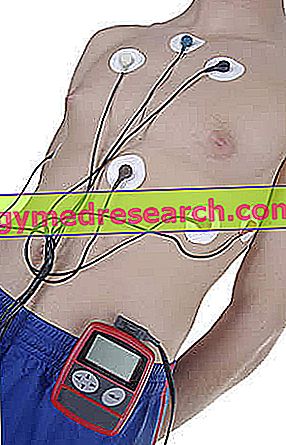
The electrocardiogram according to Holter is a type of electrocardiogram that, thanks to the use of a portable electrocardiograph, allows to monitor the cardiac function over a certain period of time, generally 24-48 hours .
The idea of creating a portable electrocardiograph, which records cardiac function for a certain number of consecutive hours, stems from the need to "capture" those discontinuous, sporadic-appearing arrhythmias, which an ECG at rest finds difficult to detect.
The general task of a physician's assistant is to install the portable electrocardiograph as a simple, fast and painless procedure that involves applying the recording electrodes (only) to the chest. Electrodes for a Holter electrocardiogram are metal plates with an adhesive portion.
From a strictly procedural point of view, the electrocardiogram according to Holter can be divided into two consecutive phases:
- The recording phase of the rhythm and electrical activity of the heart . It is the first of the two phases and goes from when the medical assistant installs and operates the portable electrocardiograph to when the same assistant or a colleague removes it.
In this phase, the instrument records and saves the cardiac function of the patient in an internal memory.
- The graphic translation phase of what was recorded in the previous phase . It is, in fact, the phase dedicated to the creation of the path with its characteristic waves.
It is up to the "usual" medical assistant or a cardiologist, who extrapolate the data from the portable electrocardiograph through a specific computerized device.
The interpretation of the resulting route is, of course, up to the cardiologist.
During the registration phase, the patient can continue to carry out his normal daily activities, clearly making sure not to bump the device and not to disconnect the electrodes.
Curiosity: in some very special cases, the dynamic electrocardiogram according to Holter can last up to 7 (seven) days.

The effort electrocardiogram involves recording the heart function of an individual, while the latter is performing a certain intensity exercise or - but more rarely - after taking a pharmacological substance that acts on the heart and causes the same effects physical exercise.
The purpose of the effort electrocardiogram is to see the behavior of the heart during physical exertion : how the heart rate varies, that heart problems can cause the body's greatest demand for blood, etc.
As in the two previous cases, the installation of the electrodes - which usually have the appearance of adhesive plasters - is the responsibility of a medical assistant.
The area of application of the recording elements is only the thorax, since the involvement of other anatomical areas would prevent the patient from moving with ease during exercise.
The classic physical exercises planned during an exercise electrocardiogram are: walking on a treadmill or pedaling on an exercise bike .
The duration of an exercise electrocardiogram, from when the patient enters the doctor's office to when the recording ends, is a few tens of minutes .
risks
The electrocardiogram is a safe and non-invasive procedure, the main drawback of which is the possibility that the removal of the electrodes will cause redness and swelling of the skin (obviously in the area of application).
It should be pointed out that the possible onset of a cardiac complication during an effort electrocardiogram is due to physical exercise and not to the electrocardiograph.
Results and graphs
The electrocardiogram allows to identify with precision the alterations of the cardiac rhythm, which may arise due to an altered conduction of the nerve impulse through the myocardium or due to the suffering of the heart such as a myocardial infarction or cardiomyopathy.
In the next sub-chapters, readers will be able to appreciate the electrocardiographic traces of some of the best-known morbid conditions of the heart.
Clearly, in order to understand the peculiarities of these tracks, it is also necessary to report the outcome of an electrocardiogram carried out by a person who is healthy from a cardiological point of view.
Please note: readers are informed that the electrocardiographic traces shown below come from the website lifeinthefastlane.com. By clicking on the relevant images it is possible to enlarge them in relation to the screen size of your device.
NORMAL ELECTROCARDIOGRAM
As emerges from the figure below, the electrocardiographic trace of a healthy person has 5 characteristic waves, identified with the capital letters P, Q, R, S and T.
- Wave P : represents the contraction of the atriums of the heart. In technical jargon, the doctors define it as the wave of depolarization of the atria .
The P wave lasts on average 0.08 seconds (but can vary from 0.05 seconds to 0.12); if it lasts 0, 08 seconds, it means that it covers 2 small squares on the sheet of graph paper.
Immediately after the P wave, there is a rectilinear stretch that ends at the Q, R and S waves and which is called the PR interval . The PR interval expresses the time it takes for the depolarization wave to propagate from the atrial sinus node along part of the electrical conduction system of the heart, present on the myocardium (specifically, atrioventricular node and His bundle).
The PR interval has a duration that varies between 0.16 seconds and 0.2 seconds, therefore it covers from 4 to 5 small squares.
- Q, R and S waves: together, these waves form the so-called QRS complex . The QRS complex represents the contraction of the ventricles and, in technical jargon, takes on the name of ventricular depolarization complex .
Typically, the QRS complex lasts 0.12 seconds, so it covers about 3 squares.
During contraction of the ventricles, relaxation of the atria takes place, previously contracted. In medical language, this relaxation is known as repolarization of the atria or rest atriums .
- Wave T : expresses the relaxation of the ventricles. In medical parlance, this relaxation takes the name of repolarization of the ventricles or return of the ventricles to rest .
After the T wave, there is a second horizontal tract, which ends at a subsequent P wave. The subsequent P wave represents the beginning of a new cycle of depolarization and repolarization of the atria and ventricles.
Taken together, the P, Q, R, S and T waves constitute the so-called PQRST complex .
Cardiologists call the interval between two PQRST complexes with the term " RR interval ". The RR interval corresponds to a cardiac cycle .
The choice of entrusting the R waves of two consecutive PQRST complexes with the task of identifying the beginning and end of a cardiac cycle is due to the fact that, as can be seen from the underlying trace, the R wave is particularly evident.
 |
| Standard duration of the intervals of a normal electrocardiogram: |
|
Curiosity: electrocardiogram of a deceased person
The trace resulting from an electrocardiogram performed on a deceased, whose heart has stopped beating, appears as a straight line, devoid of any wave

Figure: normal electrocardiogram.
ECG ATRIAL FIBRILLATION
Atrial fibrillation is an arrhythmia that makes the heart beat very fast and irregular. It may have the characteristics of a sporadic phenomenon or a chronic phenomenon. If it is sporadic, it is generally also very intense; if instead it is chronic, it is usually of reduced intensity.
To cause an atrial fibrillation is an abnormal generation of impulses that contract the atrums of the heart. This anomalous generation, in fact, causes the walls of the atrial cavities to undergo continuous and incessant stresses.
During an atrial fibrillation, the atria have a contraction frequency of approximately 350-400 beats per minute. This increased contractile frequency of the atria has repercussions on the ventricles, also strongly altering their frequency of contraction.
The electrocardiogram of a person with atrial fibrillation has the following characteristics:
- Absence of P waves. This denotes the atrial contraction defect, typical of atrial fibrillation.
- Irregular straight stretches.
- QRS complexes of irregular shape.

ECG FLUTTER ATRIAL
Atrial flutter is an alteration of the heart rhythm that is located in the atrium, such as atrial fibrillation.
Its onset coincides with a very rapid, irregular and variable intensity heart beat.
Compared to atrial fibrillation, the contraction frequency of the atria is slightly lower: in an atrial flutter, in fact, the atria contract with a frequency of about 240-300 beats per minute.
This high frequency of contraction of the atria can have repercussions on the contractile frequency of the ventricles: when it does, cardiologists speak of paroxysmal atrial flutter; when it does not, they speak of permanent atrial flutter.
The electrocardiogram of a person with atrial flutter has the following important characteristic:
- Presence of a minimum of 2 to a maximum of over 10 P waves before each QRS complex.
This succession of different P waves is called "sawtooth".
The high number of P waves indicates an atrial disturbance.

ECG INFARTO ECG
Myocardial infarction, or heart attack, is the pathological process due to which the flow of blood destined for the myocardium is inadequate to the requests, causing the death of a more or less extensive area of the heart muscle.
Often caused by atherosclerosis, this serious condition coincides with the necrosis (that is, death) of the myocardial tissue, which results in a reduction in the contractile capacity of the heart.
Among the most classic infarct symptoms, include dyspnea, chest pain, heart disease, cyanosis, hypoxia, nausea, vomiting, confusion and alterations of various kinds of heart rhythm.
There are various types of myocardial infarction. The main types are: lower myocardial infarction, anterior myocardial infarction, anterolateral myocardial infarction and posterior myocardial infarction.
Each type of heart attack determines similar anomalies on the electrocardiographic tracing, but with a different location.
Among these anomalies, the most characteristic are:
- Presence of very deep Q waves, with the disappearance of the respective successive R waves.
- Disappearance of the S wave, which goes to merge with the T wave. The result is a more or less rounded convexity, which cardiologists call the ST tract or ST elevation.

ECG VENTRICULAR FIBRILLATION
Ventricular fibrillation is an arrhythmia that affects the ventricles and alters the characteristics of the heartbeat in a profound manner.
In fact, the latter assumes a decidedly increased frequency and speed, becomes irregular, loses coordination, constantly changes its intensity and, finally, is ineffective from the mechanical point of view.
The presence of ventricular fibrillation affects cardiac output. Changes in cardiac output strongly expose the patient to episodes of cardiac arrest or sudden death.
The electrocardiogram of a person with ventricular fibrillation has the following characteristics:
- Waves with irregular, bizarre and random shapes ("box of worms")
- QRS and / or P wave complexes that are difficult to identify.
- Deviations of straight sections.

ECG COMPLETE ATRIOVENTRICULAR BLOCK
The complete atrioventricular block consists of an interruption, taking place between the atrium and the ventricle, of electrical signals that contract the heart. This results in a lack of synchrony between the various cardiac cavities.
The electrocardiogram of a person with complete atrioventricular block has the following characteristics:
- Absence of relationship between P waves and subsequent QRS complexes.
- Altered QRS complexes, due to abnormal conduction at the ventricular level.
- The ventricles depolarize independently of the atria.

ECG SINUS TACHYCARDY
Sinus tachycardia is an arrhythmia characterized by an increase in the frequency and speed of normal heart rhythm (or sinus rhythm). It does not involve any irregularity of beat and is perhaps the most widespread among the existing arrhythmias.
Usually, it is the consequence of events, such as intense physical exercise, strong emotion or a simple fever, after which the heart rate returns to normal.
Much more rarely, it is the result of severe heart disease or anemia.
The electrocardiogram of a person with sinus tachycardia has the following characteristics:
- P waves with a frequency exceeding 100 beats per minute. Remember that the normal heart rate is between 60 and 100 beats per minute.
- RR interval much shorter than normal, in terms of squares on the graph paper.
- Faster but more regular rhythm.

bradycardia
Sinus bradycardia is a reduction in normal heart rate (sinus rhythm), without any irregularity in the heartbeat.
A variety of conditions / circumstances can develop into a state of sinus bradycardia, including: nocturnal sleep, good physical condition, hypothermia, hypothyroidism, digitalis, beta-blockers, calcium channel blockers or quinidine, diphtheria, rheumatic fever, etc.
The electrocardiogram of a person with sinus bradycardia has the following characteristics:
- P waves with frequency lower than 60 beats per minute (NB: in the paired graph the frequency of the P waves is 45 beats per minute).
- Slower but regular rhythm.
- RR interval much longer than normal, in terms of squares on the graph paper.

ECG LONG QT SYNDROME
Long QT syndrome is a rare cardiac condition, which leads to an increase in the repolarisation times of the ventricles. In other words, those with long QT syndrome have a heart whose ventricles take longer than usual to relax and prepare for another contraction.
This delay in the repolarization of the ventricles favors the appearance of syncope, convulsions and severe cardiac arrhythmias, such as ventricular fibrillation.
The long QT syndrome owes its particular name to the characteristic electrocardiogram that people carry: in the electrocardiographic tracing of these subjects, the QT interval lasts for more than 0.42 seconds, which represent the maximum threshold of normality.

ECG IN BEARER OF VENTRICULAR PACEMAKER
A pacemaker is a small electronic device, capable of normalizing, through the release of electrical impulses, the cardiac contractions of people with a heart whose rhythm is too slow, too fast, or irregular.
Implanted just below the clavicle, a generic pacemaker includes these components: a pulse generator, enclosed within a metal container, and one or more cables called leads.
Leads represent the elements for conducting electrical impulses, produced in the generator, at the heart.
Depending on the number and location of the leads, a pacemaker can be: single chamber, bicameral and biventricular.
The electrocardiogram of a person with a single-chamber ventricular pacemaker has the following characteristics:
- Absence of the P wave, because it changes the generating center of electrical impulses (it is no longer the atrial sine node but the pacemaker).
- Presence of a small tip ( spike ) near the QRS complex.
- Wider QRS complexes, compared to the QRS complexes of a normal electrocardiogram.