Anatomical description
Anatomy and Physiology of the lower limb veins
The veins are blood vessels with a wall formed of three layers which, from the inside to the outside, are respectively:- The Intima tunic, covered with a single layer of extremely flat epithelial cells called endothelial cells;
- The Medium cassock, middle muscular layer, thinner than that of the arteries;
- The cassock Adventitia, the most external, formed by connective tissue (supporting collagen and elastin).
The venous system of the lower limbs is composed of three elements: the deep venous system, the superficial venous system and that of the perforators, which connect the superficial system with the deep one (and not vice versa, because in a continent the venous blood it goes from the superficial to the deep system, and also at the level of the perforators the venous flow follows this rule).
From the deep circle, then, the blood returns upwards, to the right heart, because the calf and thigh muscles, contracting during movement, "squeeze" the veins of the deep circle bringing the blood upwards, against the force of gravity.
All this is favored by many valves, which are found in all the veins of the lower limbs, and which, if fully functional, open when blood arrives and close immediately after its passage, preventing its reflux backwards., downward.
Veins and Circulation
Role of veins in blood circulation
In our cardiovascular system, venous blood is rich in carbon dioxide and waste substances, and goes from the periphery to the right heart, which will then take it to the lung to purify it from carbon dioxide and supply it with oxygen. At this point the blood becomes arterial, rich in oxygen and nutrients, and from the lung is taken to the left heart, from where it will then be distributed to the periphery).
In the lower limbs we can recognize two large superficial venous districts, which are the GrandeSafena vein (which originates from the Femoral vein) and the Piccola Safena vein. The Great Safena drains into the deep veins much of the superficial circulation of the leg and thigh, while the Small Safena essentially drains the posterior region of the leg always in the deep veins of the lower limb.
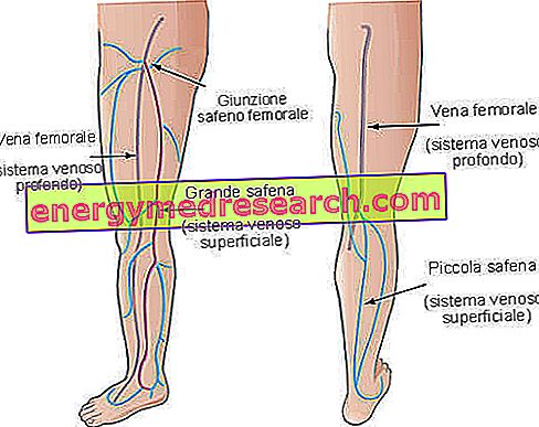
The Safene have a relationship with the deep venous systems through the perforating veins, which number about 150 in number.
During the standing (orthostatism) the haematic (sanguine) column exerts, at the level of the foot, a pressure which in the adult corresponds to an average value of 80-100 centimeters of water (which is a unit of measurement of pressure) . When physical movement begins, such as walking (for example, walking), the blood passes from the superficial venous circulation to the deep and, thanks to the pump propulsion mechanism of the leg and thigh muscles, an emptying of the deep venous circle occurs, with progressive fall in blood pressure to about 20 centimeters of water; after stopping walking, the pressure values slowly tend to recover, in about 30 seconds.
It is very important, to understand the varicose pathology, to understand what are the haemodynamic factors that come into play in the venous return. They are:
- Vis a latere, back, front, or the strength of the musculature from the side, from behind and from the front in rest conditions (tonic contractions);
- Muscle pump during movement (phasic contractions);
- Venous tone, or that ever-present degree of slight contraction of the vein wall regulated by the vegetative nervous system (ie the one that regulates many functions that do not depend on our will