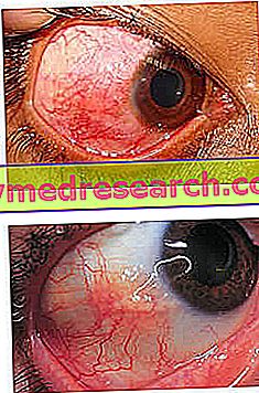
What is Episclerite?
Episcleritis is a self-limiting inflammatory disease that affects episcleral tissue.
The so-called "white of the eye", or more correctly sclera, is a fibrous membrane that externally covers most of the eyeball (approximately 5/6 of its surface).

The sclera is formed by two layers: the outermost one is called episclera and is rich in connective tissue and blood vessels. The innermost layer is instead the sclera proper, formed by a loose connective tissue.
The sclera is covered externally by the conjunctiva; in the anterior part of the eyeball is bordered by the cornea, while posteriorly it lets the optic nerve pass.
Episcleritis typically occurs with generalized or circumscribed redness of the eye, associated with mild eye pain in the absence of secretions and visual problems. Usually, the condition is idiopathic, so its cause remains unknown. In other cases, episcleritis may be associated with connective or systemic diseases. Recurrent episodes are common.
The therapy is symptomatic and includes the use of lubricating eye drops. The most severe cases can be treated with topical corticosteroids or oral anti-inflammatory drugs (NSAIDs).
Causes
The episclera is a thin layer of tissue, located between the conjunctiva and the sclera. The redness of the eyes associated with episcleritis is due to congestion of episcleral blood vessels, which extend radially.
Generally, there is no uveitis or thickening of the sclera. The disease is often idiopathic and an identifiable cause is confirmed only in one third of cases.
Episcleritis may be associated with an inflammatory, rheumatic or systemic condition such as:
- Vasculitic systemic diseases: nodular polyarthritis and Wegener's granulomatosis;
- Connective tissue diseases: rheumatoid arthritis and systemic lupus erythematosus;
- Hyperuricemia and gout;
- Inflammatory bowel diseases: ulcerative colitis and Crohn's disease;
- Rosacea, atopy, lymphoma and thyroid orbitopathy (pathology of the eye orbit of thyroid origin).
Infectious causes are less common; it is the case of: Herpes zoster, Herpes simplex, Lyme disease, syphilis, hepatitis B and brucellosis. Contact with chemicals or a foreign body can also cause episcleritis.
Rarely, the condition is caused by scleritis, a severe inflammation that occurs throughout the thickness of the sclera.
Episcleritis occurs more commonly in young adults, especially in women. However, there are no specific risk factors for the disease.
Signs and symptoms
To learn more: Episclerite symptoms
Episcleritis symptoms include mild eye pain, hyperemia of the globe, irritation and watery eyes. Furthermore, photophobia, eyelid edema and conjunctival chemosis may be present. The ocular secretions are absent and the vision is unaffected. The onset is acute or gradual, widespread or localized.
There are two main types of episcleritis:
- Simple episcleritis : it is a recurrent inflammation, but self-limiting, which affects the episclera partially (simple sectorial episcleritis) or diffuse (simple diffused episcleritis).

- Nodular episcleritis : involves a well-circumscribed area of the episclera and is characterized by the presence of a small raised and translucent nodule in the inflamed area. In nodular episcleritis, attacks are self-limiting, but tend to last longer.
Episcleritis is distinguished from conjunctivitis by hyperaemia localized to a limited area of the globe and by less profuse tearing. Furthermore, pain is less severe than scleritis and photophobia is less than uveitis. Episcleritis does not cause the presence of cells or blood spills in the anterior chamber of the eye. Rarely, some cases may progress to scleritis.
Diagnosis
The diagnosis of episcleritis is clinical and is based on the anamnesis and physical examination. Further investigations are necessary for some patients in order to identify a possible basic medical condition.
Episcleritis can be differentiated from scleritis by instilling phenylephrine-based eye drops. This substance causes the bleaching of the superficial and conjunctival episcleral vascular network, but leaves the underlying scleral blood vessels undisturbed. If the redness of a patient's eyes improves after the application of phenylephrine, the diagnosis of episcleritis can be confirmed.
The examination with the slit lamp allows to distinguish the nodular form from the sclerite. Furthermore, it is important to note that the nodule is raised and freely movable with respect to the underlying scleral tissue.
Treatment
Often, treatment is not necessary, as episcleritis is a self-limiting condition. Most cases resolve within 7-10 days, but patients should be aware that the episodes may recur in the same or the other eye. Nodular episcleritis is more aggressive and takes longer to heal (about 5-6 weeks).
Artificial tears can be used to relieve irritation. Severe or chronic / recurrent cases can be treated with corticosteroids and non-steroidal anti-inflammatory drugs for topical or oral use. These measures help reduce inflammation and accelerate recovery, but there are some risks associated with the use of steroid eye drops. The patient must therefore be carefully monitored by the doctor during therapy.
In general, episcleritis does not cause complications in the eye structures; there may occasionally be corneal involvement (in the form of inflammatory cell infiltration) or edema; in addition, recurrent attacks over a period of years can cause mild scleral thinning.


