The joints are anatomical structures, sometimes complex, which put two or more bones in mutual contact. To avoid degenerative phenomena due to wear, in most cases it is a non-direct contact, but mediated by fibrous or cartilaginous and / or liquid tissue.
The joints of the human body are very numerous, there are an average of 360, and structurally very different from each other. This diversification reflects the type of function required for that particular joint. Taken together, the task of the joints is to hold the various bone segments together, so that the skeleton can fulfill its function of support, mobility and protection.
CLASSIFICATION OF JOINTS ON STRUCTURAL BASE
The joints are subdivided, from the structural point of view, into:
- fibrous joints : the bones are joined by fibrous tissue;
- cartilaginous joints : the bones are linked by cartilage;
- Synovial joints : the bones are separated by a cavity, as well as being tied up by means of structures that we will describe better later.
However, the best known subdivision is on a functional basis. The bones of the human skeleton are in fact connected by means of joints to which movements of various types and degrees are allowed. We speak, then, of motionless joints (synarthrosis), semi-mobile (amphiarthrosis) and mobile (diarthrosis).
CLASSIFICATION OF JOINTS ON FUNCTIONAL BASE
The joints are divided, from the functional point of view, into:
- motionless joints or synarthrosis: they bind tightly the bone heads, like a closed zipper, so as to prevent their movements.
- Hypomobilized joints or amphiarthrosis : they bind two articular surfaces, covered by cartilage, through interosseous ligaments; a fibrocartilaginous disk is interposed between the two surfaces which allows only limited movements. In the vertebrae, for example, flat bony surfaces are joined by a cartilaginous interosseous disc that acts as a shock absorber.
- Mobile joints or diarthrosis : allow a wide range of movement, in one or more directions of space (knee, shoulder, fingers ...)
The structure of a joint influences the degree of mobility:
Functional name | Structural name | Degree of movement | Example |
sinartrosi | fibrous | fixed | skull |
anfiartrosi | cartilaginous | little mobile | vertebrae |
diarthrosis | synovial | very mobile | shoulder |
Synarthroses (immobile joints) are divided into:
- Synostosis: the degree of movement is zero, since they join the joints through bone tissue (as in the adult's skull).
- Synchronous: the degree of movement is scarce, since they join the joints through dense cartilaginous tissue (like the first ribs of the sternum).
- Syndesmosis or sinfimbrosi: the degree of movement is limited, since they are held together by fibrous connective tissue (such as the pubic symphysis).
The movable or semi-mobile joints differ in the shape and the allowed movements. In this respect there are slightly different classifications. One of these is the subdivision of diarthrosis based on the differences in the shape of the articular surfaces:
Artrodia | |
| Permitted movements: simple scrolling | |
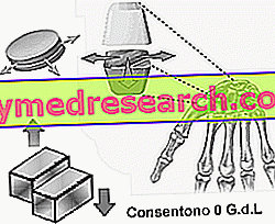 | The arthrodias, which combine the bones of the carpus in the hand and the tarsus in the foot, allow only small sliding movements. Flat bone surfaces simply slide over one another to allow for minimal movement. The carpal bones, for example, slide between them during hand movements. They have the task of amortizing the shocks. Further examples: cost-vertebral joints. |
Trocleoarthritis (angular ginglimo) | |
| Permitted movements: inflection / extension | |
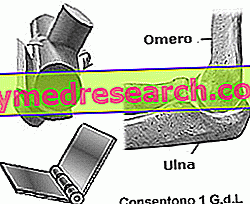 | The articular surfaces that face each other have the shape of a cylinder segment, one of which, with a concave throat (trochlea), fits into the convex face of the other. The axes of the cylinders are orthogonal (at right angles) . The movement takes place in a plane according to a single axis (uniaxial), like a door in the hinge. Example: elbow, knee |
Trochoid (lateral / parallel ginglimo) | |
| Permitted movements: pronation and supination | |
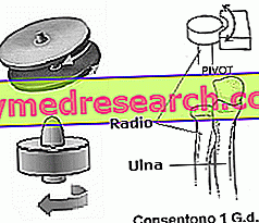 | The two articular surfaces have the shape of a cylinder segment, one of which, with a concave throat (trochlea), fits into the convex face of the other. The axes of the cylinders are parallel. It is a uniaxial articulation. Example: between the capital of the radius and the ulna (proximal radio-ulnar joint). |
A Sella or Pedartrosi | |
| Permitted movements: extension flexion, abduction adduction, circling | |
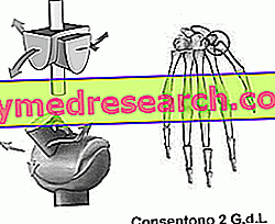 | They are joints consisting of two surfaces each having two curvatures, one concave and the other convex. Example: between the carpus and the metacarpus of the thumb; between the sternum and the clavicle. |
Condilartrosi | |
| Permitted movements: extension flexion, abduction adduction, circling | |
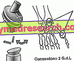 | They are joints consisting of two ellipsoidal surfaces, of which one full (condyle) is housed in another convex (condylar cavity). Example: between the radio and the carpus; between the metacarpus and the phalanges; the knee joint; temporomandibular joint. |
ball and socket joint | |
| Permitted movements: extension flexion, adduction abduction, circumduction, intra and external rotation | |
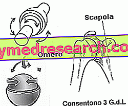 | They are joints consisting of an articular head similar to a full sphere (head) housed in an articular cavity in the shape of a hollow sphere. The movements are performed along all three fundamental axes (sagittal, transverse and vertical) They are the most mobile joints in the human body. Example: hip joint (Coxofemoral); articulation between scapula and humerus (scapula-humeral). |


