Anatomy and Functions
Carotid arteries are two large blood vessels located at the sides of the neck; together with the vertebral arteries, the carotids with their numerous branches supply the head and neck, transporting oxygen-rich blood from the heart to the brain and to the facial structures .
The left common carotid artery derives directly from the aortic arch, while the right one arises from the innominate (or anonymous) artery.
Anatomically, viascuna carotid is distinguishable in:
- Common carotid artery ;
- Internal carotid artery ;
- External carotid artery
The common carotid arteries ascend deeply into the neck and divide at the level of the larynx (Adam's apple) in an external carotid artery and an internal one.
- The external carotid arteries supply the following structures: neck, pharynx, esophagus, larynx, mandible, scalp and face.
- The internal carotid arteries, on the other hand, enter the skull at the level of the carotid holes of the temporal bones, bringing blood to the brain. From here they go up to the level of the optic nerve, where they divide into three branches: ophthalmic artery (vasculature of the eye), anterior cerebral artery (irrigates the frontal and parietal lobes of the brain) and middle cerebral artery (brings blood to the midbrain and to the lateral structures of the cerebral hemispheres).
The carotid sinus, located at the base of the internal carotid artery,


The brain is extremely sensitive to changes in vascular supply, so that an interruption of circulation for a few seconds will produce unconsciousness, while after about four minutes the brain damage will be permanent. These circulatory crises are rare, as the blood can reach the brain even through the vertebral arteries .
Internal carotids generally supply blood to the anterior half of the brain, while the rest of the brain receives blood from the vertebral arteries. However, this distribution can easily change: the internal carotid arteries and a portion of the vertebral artery (ie the basilar artery) are interconnected by the circle of Willis, an anastomotic ring circuit, which surrounds the pituitary gland. Thanks to this cerebral arterial circulation, the possibility of a serious interruption of the vascular supply to the brain is reduced.
Carotid stenosis
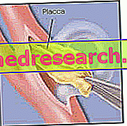
Carotid arteries, like any other healthy artery, are flexible and have smooth inner walls. Following a process called atherosclerosis, their walls may however undergo a progressive stiffening accompanied by the reduction of the inner lumen; this phenomenon is caused by the gradual accumulation of deposits ( atheromatous plaques ) consisting of fats, proteins, fibrous tissue and other cellular debris. Over time, these plaques can form a large mass that reduces the internal diameter of the artery, limiting blood flow (this is called carotid stenosis). The atheromatous deposits are formed above all in the carotid sinus, that is at the level of the bifurcation which divides the common carotid artery into the internal and external carotid artery.
The obstructive disease of the carotid artery develops slowly and often goes unnoticed: the first indication of the presence of the atheroma can already be very serious, such as the appearance of a cerebral stroke or a transient ischemic attack ( TIA ).
The treatment of carotid stenosis aims to reduce the risk of significantly reducing blood supply to the brain, removing atheromatous plaque and controlling blood clotting (to prevent thromboembolic stroke).
Symptoms
In the early stages, the obstructive carotid artery disease often produces no sign or symptom. Stenosis may become evident only when it becomes severe enough to deprive the brain of blood, causing a stroke or transient ischemic attack (TIA), both signs of early warning for a future apoplectic attack.
Signs and symptoms of a transient ischemic attack or stroke may include:
- Sudden numbness of the face or weakness in the limbs, often on only one side of the body;
- Inability to move one or more limbs;
- Difficulty speaking and understanding;
- Sudden difficulty in vision, in one or both eyes;
- Vertigo and loss of balance;
- A sudden, severe headache, with no known cause.
Although the signs and symptoms last only a short time (sometimes less than an hour) it is possible that the patient has experienced a TIA. If any of these manifestations occur it is important to seek emergency care, to increase the chances that the carotid artery disease is detected and treated promptly, before a disabling stroke occurs. It is not excluded that a TIA may be due to the lack of blood flow also in other vessels: the doctor is able to establish which tests are necessary to ascertain the condition.
Complications of carotid stenosis
The most serious complication of the obstructive disease of the carotid artery is stroke, as it can cause permanent brain damage and, in severe cases, can be fatal.
There are three different ways in which the presence of an atheromatous plaque increases the risk that this may occur:
- Reduced blood flow . As a result of atherosclerosis, the lumen of the carotid artery may undergo such a reduction, that the supply of blood is not sufficient to reach some parts of the brain. The atheroma can eventually completely occlude the artery.
- Plaque rupture . A piece of atheromatous plaque can break and break off, traveling to the smallest arteries in the brain. The fragment can remain blocked in one of these cerebral arteries, creating an obstruction that blocks the flow of blood to the area of the brain that the blood vessel irrigates.
- Blood clot obstruction . Some plaques are prone to cracking and deforming the artery wall. When this happens, the body reacts as a lesion, sending platelets locally, to facilitate the coagulation process. In this process, a large blood clot can develop and block or slow the flow of blood through a carotid or cerebral artery, leading to a stroke.
Risk factors
The combination of several factors can increase the risk of injury, the formation of plaques and the onset of carotid stenosis are:
- High pressure. Arterial hypertension is an important risk factor for obstructive carotid artery disease. Excessive pressure on the walls of the arteries can weaken them and make them more vulnerable to damage.
- Smoke. Nicotine can irritate the inner lining of the arteries. In addition, it increases heart rate and blood pressure.
- Age. Older people are more likely to be affected by carotid stenosis, as with age, arteries tend to be less elastic.
- Abnormal levels of fat in the blood. High levels of low density lipoproteins (LDL, the "bad" cholesterol) and triglycerides in the blood favor the accumulation of atheromatous plaques.
- Diabetes. The pathology not only influences the ability to manage glucose appropriately, but also the ability to efficiently process fats, placing the patient at greater risk of hypertension and atherosclerosis.
- Obesity. The extra pounds contribute to other risk factors, such as hypertension, cardiovascular disease and diabetes.
- Heredity. If the patient has a family history of atherosclerosis or coronary heart disease, he has an increased risk of developing these diseases.
- Physical inactivity. Lack of regular exercise predisposes to a number of conditions, including hypertension, diabetes and obesity.
Diagnosis
In addition to considering the complete history, the presence of risk factors and any signs or symptoms, the doctor can carry out various tests to evaluate the health of the carotid arteries:
- Physical examination. The doctor can auscultate the carotid artery by placing a stethoscope at neck level to detect a sound similar to a "suction", characteristic of the turbulent blood flow caused by atherosclerosis. The doctor can perform a neurological evaluation to check the patient's physical and mental status, such as stamina, memory and speech.
One or more diagnostic tests can be performed to evaluate the narrowing of a carotid artery:
- Doppler ultrasound: non-invasive test that uses reflex sound waves to assess blood flow through the blood vessel and check for the presence of a possible stenosis. The ultrasound probe is placed on the neck, at the level of the carotid arteries. Doppler ultrasound reveals how blood flows through the artery and to what extent the contribution is reduced (carotid stenosis less than 0-49%, moderate 50-69% and severe 70-99%, until complete obstruction).
- Angio-CT (CTA): provides detailed images of the anatomical structures of the neck and brain. The investigation involves the injection of a contrast agent into the blood stream, so as to highlight the abnormalities of blood vessels (via angiography) and soft tissues (by computerized tomography). CTA allows doctors to visualize the narrow carotid artery and determine the pathological degree of stenosis.
- Angiography using magnetic resonance imaging (MRA): like the CTA, this imaging test uses a contrast agent to highlight the arteries that supply the neck and brain. The magnetic field and radio waves are used to create three-dimensional images.
- Magnetic resonance imaging (MRI): allows visualization of brain tissue to detect stroke or other abnormalities early.
- Cerebral angiography: it is a minimally invasive test that uses X-rays and a contrast agent injected into the arteries, through a catheter inserted directly into the carotids. Cerebral angiography allows doctors to visualize in detail all the arteries that supply the brain.
Imaging can also reveal evidence of multiple transient ischemic attacks. Doctors can define the diagnosis of carotid stenosis, if evidence shows that blood flow is decreased in one or both carotid arteries.
Treatments and Drugs
The goal from therapy is to reduce the risk of stroke. Treatment options for carotid stenosis vary depending on the severity of arterial narrowing and whether symptoms occur (asymptomatic).
Mild to moderate carotid stenosis
- Lifestyle change. Changes in behavior can help reduce pressure on the carotid artery and slow the progression of atherosclerosis. Such changes include quitting smoking, losing weight, drinking alcohol in moderation, eating healthy foods, reducing the amount of salt and practicing regular exercise.
- Manage chronic conditions. With the doctor, it is possible to establish a therapeutic plan to deal properly with specific chronic conditions, such as high blood pressure, excess weight or diabetes, which can also produce pathological effects on the carotid arteries.
- Drugs. Asymptomatic patients or patients with low-grade carotid stenosis are treated with drugs. Your doctor may prescribe a platelet antiplatelet (such as aspirin, ticlopidine, clopidogrel), to be taken daily to thin the blood and prevent the formation of dangerous blood clots. Antihypertensive drugs can also be recommended to control and regulate blood pressure (ACE inhibitors, angiotensin blockers, beta-blockers, calcium channel blockers, etc.) and statins to lower cholesterol and help reduce plaque formation in the atherosclerosis. Statins can reduce "bad" LDL cholesterol by an average of 25-30% when combined with a low-calorie, low-cholesterol diet.
Severe carotid obstruction
When you have severe stenosis, especially if the patient has already had a TIA or stroke related to the occlusion, it is best to proceed surgically by clearing the artery from the atheromatous plaque.
- Carotid endarterectomy. This surgical procedure is the most common treatment to remove the atheroma in the presence of a severe clinical picture. The operation is performed under general anesthesia.

- Carotid angioplasty and stenting. Carotid stent placement is a less invasive procedure than carotid endarterectomy, as it does not involve an incision in the neck. Carotid angioplasty with stent insertion allows good results to be obtained in the short term, and is typically indicated for patients who: 1) have a moderate-severe degree of carotid stenosis; 2) suffer from other medical conditions that increase the risk of surgical complications; 3) show a repeat offense. In carotid angioplasty, a catheter is inserted into the carotid area blocked in the neck. A specially designed filter on a guide wire (called an embolic protection device) is inserted to collect any debris that may detach from the plate during the procedure. Once in position, a small balloon is inflated at the end of the catheter for a few seconds, in order to open or enlarge the artery. A stent is inserted permanently to form a scaffold, which helps to support the walls of the arteries and to keep the lumen of the carotid artery open. The balloon is then deflated and the catheter and filter are removed. After several weeks, the artery heals around the stent. As with carotid endarterectomy, there are some risks associated with the procedure (stroke or death). Stenting will therefore be recommended only in the event of severe stenosis.

Recovery from surgical procedures generally requires a short hospital stay. Patients often return to normal activities within a week or two. After a carotid endarterectomy, the stenosis can recur and is often related to the progression of atherosclerotic disease. These new plaques can be treated by repeating the surgery.


