Generality
Osteonecrosis means death of bone.
Contrary to what many believe, bone is a living tissue that needs blood and oxygen; if these fail, the cells that compose it undergo premature death and osteonecrosis occurs.

Figure: X-ray of the shoulder joint; in the image to the right the process of osteonecrosis affecting the head of the humerus and the glenoid cavity of the scapula is evident. From the site: www.drlox.com
It can lead to local fractures and, in the most severe cases, the collapse of the affected bone.
The factors that can trigger a process of osteonecrosis are numerous, including joint trauma, abuse of corticosteroid drugs, alcoholism, sickle cell anemia, etc.
In general, the epiphyses of the long bones are affected, that is the terminal bones like the femur, the tibia or the humerus involved in the respective joints.
Pain and poor joint function are the main symptoms of osteonecrosis.
The therapy consists of various remedies, both conservative and surgical. To avoid complications, it is good to take prompt action.
What is osteonecrosis?
Osteonecrosis is the death of bone tissue, due to a missed or insufficient blood supply. Also known as avascular necrosis, bone necrosis or bone infarction, it results in the appearance of tiny fractures in the affected bone tract; in the most serious cases, osteonecrosis can even determine bone collapse .
Epidemiology
Osteonecrosis can affect anyone, however, according to some statistical surveys, it is more frequent among male individuals aged between 30 and 60 years.
Causes
The flow of blood to a certain portion of bone can be affected by:
- A joint injury located nearby . The most frequent injuries to joints that result in bone necrosis are knee sprains, shoulder or hip dislocations, ankle sprains, etc.
- Radiotherapy treatment for cancer (radiotherapy) . Ionizing radiation used to treat tumors can have unpleasant side effects; among these, the weakening of the bones and the damage to the blood vessels that supply the bone tissue and keep it alive are also included.
- Sickle cell anemia . In this disease the red blood cells have a particular shape, which makes the blood flow inside the smaller blood vessels abnormal. This results in poor blood circulation and a lack of nourishment of some tissues, including bone.
- Alcohol abuse . Fat deposits are created in the blood vessels of those who drink a lot of alcohol, which prevent the blood from flowing freely. In the long run, these deposits can completely block the caliber of the vessels and cause episodes of osteonecrosis.
- Prolonged and / or high doses of corticosteroid and bisphosphonate drugs . Corticosteroids are powerful anti-inflammatories, while bisphosphonates are medicines used in case of osteoporosis. The former, if taken in excessive doses, can cause various side effects, including fat accumulation inside the vessels and their consequent occlusion (NB: the process is very similar to what happens in case of alcohol abuse).
The latter, instead, if taken in high doses can cause a process of osteonecrosis of the jaw (NB: the pathophysiological mechanism, for bisphosphonates, has not yet been fully clarified).
Furthermore, according to some studies, they are more at risk of osteonecrosis:
- People with particular disease states, such as diabetes, AIDS, systemic lupus erythematosus, so-called decompression sickness, hypertension, Gaucher disease, arterial thrombosis, arterial embolism and rheumatoid arthritis.
- People who have had an organ transplant or are undergoing dialysis due to a serious kidney problem.
Finally, a small proportion of subjects with osteonecrosis develop the problem without any precise reason. In these situations, one speaks of idiopathic osteonecrosis .
Symptoms and Complications
To learn more: Symptoms Osteonecrosis
Very often, in the early stages, osteonecrosis is asymptomatic (ie it does not cause any obvious symptoms). Then, over time, it becomes a cause of pain and unusual fractures.
In the most serious stages (complications), the necrotic bone yields completely (collapsing).
If the death of the bone tissue also involves a (very frequent) articulation, this can degenerate in a worrying way, even losing its functionality.

Figure: head of a normal femur (left) and affected by osteonecrosis (right). The blue capillaries indicate the avascular origin of the process; also note the rarefaction of necrotic bone tissue. From the site: www.drlox.com
WHAT ARE THE MOST BEAUTIFUL BONES?
The bones most affected by osteonecrosis are the long bones of the human body, such as the femur (ie the thigh bone) and the humerus (ie the arm bone).
This is true both when the causes are traumatic, and when the patient abuses certain substances or is affected by one of the health disorders mentioned in the chapter dedicated to causes.
The epiphyses, or the terminal portions of the long bones that participate in the joints, are fractured and eventually collapsed.
WHAT ARE THE MOST AFFECTED JOINTS?
The joints most prone to osteonecrosis are the knees, the shoulders, the ankles, the wrist, the hip and the jaw.
WHEN TO REFER TO THE DOCTOR?
It is advisable to contact your doctor when a certain bone or joint is strangely painful. If osteonecrosis is diagnosed in time, the most serious complications can be avoided.
Diagnosis
To diagnose osteonecrosis, physical examination is not enough; many pathologies, in fact, present symptoms similar to those of a bone infarction. Hence the need to resort to specific instrumental examinations.
The three most practiced tests are:
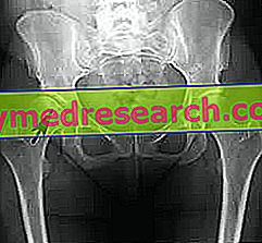
- X-ray examination . It is useful in detecting the bone changes that occur in the advanced stages of osteonecrosis; at the beginning, in fact, the problem is rarely identified.
During radiographs the patient is exposed to a minimal dose of ionizing radiation.
- Bone scintigraphy . Through the injection of a radiopharmaceutical intravenously, it allows to obtain images related to the anatomy and activity of the bones. This is a very sensitive diagnostic test, because it clearly shows if there are pathological changes; however, at the same time, a non-specific examination appears, as it does not clarify the nature of the disturbances highlighted.
The quantities of radioactive substance used are low, however bone scintigraphy is not recommended during pregnancy.
A big advantage of MRI is that it is completely harmless to the patient.

THE PROBLEM OF LATE DIAGNOSIS
It often happens that osteonecrosis manifests itself with symptoms and signs only at an advanced stage, when by now the bone tissue is already partially compromised. In these cases, the diagnosis is inevitably late and this could affect the effectiveness of the treatment.
Treatment
To reduce the symptoms and slow down the progression of osteonecrosis, various drugs and some conservative treatments are available, such as simple rest and physiotherapy. However, when the bone infarction reaches a certain stage, these remedies can be ineffective or insufficient. On such occasions it is necessary to resort to more invasive, surgical treatments, such as bone decompression surgery or even the operation to insert a joint prosthesis.
PHARMACOLOGICAL THERAPY
The possible medicines prescribed for osteonecrosis cases are:
- NSAIDs, or Non-Steroidal Anti-Inflammatory Drugs . They are used to alleviate the sense of pain and to reduce the inflammatory processes that affect the affected bone. The most widely used NSAID is ibuprofen.
- Some osteoporosis drugs . Research has shown that alendronate bisphosphonate, normally used against osteoporosis, also slows the progression of bone necrosis. However, it should be remembered that the drugs belonging to the class of bisphosphonates, if given in excess, have different side effects, including osteonecrosis of the jaw.
- Cholesterol-lowering drugs . These medicines, by reducing the amount of lipids in the blood, are administered with the intent to improve blood circulation in the vessels that caused osteonecrosis.
- Anticoagulants . They prevent the formation of blood clots and allow better blood circulation, even in the vessels involved in osteonecrosis.
REST AND PHYSIOTHERAPY
Rest is essential to avoid further stressing the aching bone and / or joint. In cases of osteonecrosis affecting the hip or knee, the use of crutches is recommended for a certain time interval.
Physiotherapy, on the other hand, is especially useful when the infarct has reduced the mobility of a joint.
SURGICAL THERAPY
Surgery is used when osteonecrosis is found in very advanced stages, so it cannot be treated in any other way. Here are the surgical treatments available:
- Bone decompression . It consists in removing the diseased bone portion, in order to stimulate the remaining healthy portion to regenerate new bone.
- Bone transplantation . It consists in replacing the diseased bone tract with a piece of bone taken from a healthy part of the body (autograft) or from a donor.
- Osteotomy . It consists in dissecting the diseased bone, removing some parts in order to redistribute, on a still healthy bone portion, the weight that weighs on the necrotic area. In order to perform the osteotomy, of course, the bone must still have areas of living and strong tissue.
- Joint prosthesis . It is the intervention for the most serious cases, in which a bone collapse occurred. There are hip replacement procedures, knee prostheses etc.
Prevention
Preventing osteonecrosis due to certain health conditions is not at all easy.
However, it may be important to limit alcohol intake, to ensure that the level of cholesterol in the blood remains low and, if you are taking corticosteroids, scrupulously follow the doses recommended by your doctor.
Prognosis
Late therapy or lack of treatment make the prognosis negative, as osteonecrosis is a chronic, debilitating degenerative problem.