Introduction
Due to ethical reasons and due to the difficulty of using invasive in vivo methods, there are very few data from clinical studies performed on healthy newborns and children.

Characteristics of the Child's Plle
The baby's skin in early life is often considered an ideal cosmetic reference for adults. However, compared to that of an adult it seems to be more prone to develop certain pathological conditions, such as atopic dermatitis and contact dermatitis.
The baby's skin has high TEWL, high pH, flaking, high cellular turnover and a high water content despite the fact that the NMF value (skin hydration factor) and the concentration of surface lipids are lower than the levels found in adult skin. As a result, the function of the epidermal barrier can be inefficient, making the baby's skin susceptible to the onset of disease and vulnerable to chemical agents and microbial aggression.
Understanding the physiology of the skin of the healthy child in the first years of life is therefore necessary both from the cosmetic point of view (development of products suitable for the child's skin) and from the clinical point of view (understanding and treatment of dermatological problems).
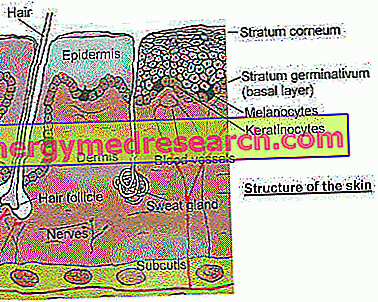
Baby Skin Structure
The skin performs many different vital functions, such as physical and immunological protection from external agents (UV radiation, microorganisms, humidity, extreme temperatures). It has a thermoregulatory, moisturizing, sensory, excretory and secretive function.
Skin development begins within the uterus during the first trimester of pregnancy and continues with functional maturation of the stratum corneum up to about the 24th week of gestational age. During the last trimester of pregnancy, the formation of the caseous paint is also observed, a protective coating of the skin, derived from sebaceous secretions and dead corneocytes, and largely composed of water, lipids and proteins. Its function is to isolate the skin of the fetus from the amniotic fluid of the uterus thus avoiding the maceration of the skin itself; moreover, it helps to make the child's intense environment change less traumatic at birth. The maturation of the skin is a gradual process and the level of maturity is a function of the gestational age. In premature infants, in fact, the function of the epidermal barrier is weaker.
- Structure of the skin microrelief: At birth, the newborn's skin is relatively rough compared to older children but becomes smoother and smoother during the first thirty days of life. The skin texture appears denser in the newborn and under the microscope small homogeneous corneal lamellae are visible in terms of size, density and distribution. The structural relationship between the epidermal lamellar islands and the underlying dermal papillae, not perceptible in adults, justifies the better hydration of the stratum corneum of the child compared to that of the adult.
- Horny layer and epidermal thickness: The thickness of the stratum corneum and that of the epidermis appear respectively 30 and 20% thinner in children between 6-24 months of age compared to the size measured in the adult. The skin is therefore more fragile in the face of external mechanical stimuli; hence the value and importance of the barrier function of the skin, the alteration of which can give rise to irritative moments characterized by transient redness and peeling, aggravated by insufficient thermoregulation capacity. Over the years the skin thickness increases until it reaches a maximum in young adults, and then gradually decreases with the aging process.

- Dermal collagen and elastin: the skin of children in the first years of life presents a derma not very thick, since the collagen fibers and the elastic fibers, although abundant, are still immature. The collagen fibers are in the upper part of the dermis less dense than in the adult and it is not possible under the microscope to distinguish the reticular dermis from the papillary dermis. The vascular and neural components are also not very organized, as well as the dermo-epidermal junctions are not yet well welded. These structural differences could, at least in part, underlie the functional differences observed between the adult's skin and that of the child.


