Generality
Breast fibroadenoma is a benign breast tumor that mainly affects young women. Of variable size, it resembles a marble and can take on a sometimes gummy consistency, sometimes stiff.
The triggers at the moment are still unclear.

Figure: a fibroadenoma in the breast. From the site: daviddarling.info
However, the major suspicions concern estrogens (female sex hormones) and the role they played during the second and third decades of life (ie between 15 and 30 years).
Most fibroadenomas are harmless and do not cause malignant breast tumors, however, to be sure of their kindness, one must undergo an accurate diagnosis.
The therapeutic treatment, when foreseen, involves a surgical removal operation.
What is breast fibroadenoma?
Breast fibroadenoma is a solid tumor of benign type, which occurs in the breasts (or mammary glands ) of young women.
On palpation, the fibroadenoma appears as a nodule with a smooth surface and variable consistency: in some cases, in fact, it can be rubbery; in others, it may be rigid; at other times, it is described as a very mobile marble.
Breast fibroadenoma is not to be considered a dangerous condition as is the malignant breast tumor, however its occurrence requires periodic monitoring, for the sole purpose of precaution.
Deepening: what is a benign tumor?
With the term of tumor, a mass of proliferating cells is identified, which is formed, in a completely anomalous way, inside a tissue.
In the case of benign tumors, the cell mass proliferates very slowly, is not infiltrative or even metastasizing (for non-infiltrative and non-metastatizing, it means that it does not invade the surrounding tissues and does not spread to the organism, as it does, instead, a malignant tumor). A malignant tumor (or cancer or carcinoma) has exactly the opposite characteristics: it grows quickly and, if not removed in time, spreads to the surrounding tissues and the rest of the body.
BREAST TYPES OF FIBROADENOMA
There are two types of breast fibroadenoma: the simple fibroadenoma and the complex fibroadenoma.
The simple fibroadenoma is a cell mass generally free of any danger, which remains stable (or even reduces) over the course of a lifetime.
The complex fibroadenoma, on the other hand, is a benign neoplasm to be kept under periodic observation, because, according to accredited scientific studies, it would seem to favor the appearance of a malignant breast tumor.
The differences between simple fibroadenoma and complex fibroadenoma also concern the internal structure: in complex fibroadenomas, in fact, cysts full of fluid and calcium deposits can be identified, both absent in simple fibroadenomas.
To know which type of fibroadenoma you suffer from, you need to undergo the appropriate diagnostic tests.
IN WHICH POINT OF THE BREAST DOES THE FIBROADENOMA DISPLAY?
The breast or mammary gland has a particular anatomy. Inside, it contains glandular tissue, adipose tissue and fibrous tissue. The glandular tissue is composed of structures called lobules, which, joining together, form the so-called lobes .

Figure: the female breast and its main structures. From the site: breastcancercare.org.uk
In the lobes, breast milk is produced, which reaches the nipple through small canals, called milk ducts .
Breast fibroadenoma is an abnormal compact mass that forms at the level of the lobules.
Epidemiology
Breast fibroadenoma is a benign tumor typical of young women, ranging in age from 15 to 30 years. According to some statistical data, in fact, starting from the age of 30, the chances of its appearance are gradually reduced, to the point that, in the post-menopausal period, they are really very few.
According to a study carried out in the United States, the annual incidence of breast fibroadenoma concerns about 10% of American women.
Causes
The precise causes, which cause a breast fibroadenoma, have not yet been fully clarified.
According to some studies, the origin seems to be linked to too high levels of estrogen, or female sex hormones . If this were the case, it would be better to explain the high incidence among young women, in which there is a considerable amount of circulating estrogens for the body, but also the low incidence among those in menopause and post-menopause, where the number of estrogen is significantly reduced.
Symptoms and Complications
Breast fibroadenoma has the following characteristics:
- To the touch, it looks like a marble or a lump
- It has well-defined and regular contours
- It can be rigid or rubbery
- It has a smooth surface
- It is usually painless
- It is of variable size
DIMENSIONS
The size of a breast fibroadenoma can vary from person to person.
Some women have fibroadenomas of small diameter, equal to 1-3 centimeters; others, instead, develop much more voluminous cell masses, with a diameter that can reach even 5-6 centimeters.
Small fibroadenomas are generally of the simple type, while large fibroadenomas can belong to both the simple and the complex type.
In menopause, a definitive regression of fibroadenoma can occur, which leads to the latter becoming almost imperceptible to touch and sight; vice versa, during a pregnancy, the volume of the tumor may temporarily increase, possibly due to a change in hormone levels.
HOW MANY FIBROADENOMES IN THE BREAST CAN BE FORMED?
On a woman's breast, one or more fibroadenomas may appear. When there are more than one, only one or both can be affected.
If the cellular masses are of the simple type (ie they do not contain cysts and / or calcium deposits), the appearance of more fibroadenomas in the breast is completely random and has no particular meanings.
WHEN TO REFER TO THE DOCTOR?
At the onset of a fibroadenoma in the breast or in front of its possible enlargement, it is always advisable to consult your doctor and undergo precautionary clinical tests. Only in this way, in fact, it is possible to discover if the benign tumor in question is of the simple type or of the complex type.
COMPLICATIONS
The presence of simple breast fibroadenomas is generally considered harmless.
Therefore, the only documentable complications are those related to a possible evolution of a complex fibroadenoma in a malignant breast tumor.
Diagnosis
To establish a correct diagnosis of breast fibroadenoma, we start, as often happens, from an objective examination; after which, based on what emerges from the latter and the patient's age, instrumental examinations (mammography and breast ultrasound) and a cytological analysis (ie cells) of the tumor mass are used.
Below is an overview of the diagnostic pathway that is generally put into practice in case of suspected breast fibroadenoma:
- Physical examination . The doctor carefully analyzes the breast affected by the suspicious nodule and tries to understand its shape and size. As mentioned above, a breast fibroadenoma resembles a marble and has, on the touch, a smooth surface and a sometimes gummy, sometimes stiff consistency. Discrete fibroadenomas are easily visible even with the naked eye; those of smaller dimensions are better evident to the touch.
- Mammography . Mammography is an X-ray examination, which provides clear images of the area with the suspicious nodule. A breast fibroadenoma is presented as a mass without asperity (therefore smooth), with defined contours, of a rounded shape and, finally, distinct from the rest of the glandular tissue. This last aspect, or the fact that the tumor is isolated, is particularly important, as it is what characterizes non-infiltrating breast tumors.
Mammography is the ideal test to track breast fibroadenomas in women over 30 years.
Since it is an X-ray examination, mammography involves the use of harmful ionizing radiation.
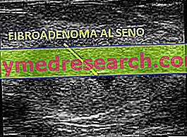
- Breast ultrasound . The ultrasound examination allows to visualize, on a monitor and in real time, the structure of the suspicious nodule.

Unlike mammography, ultrasound does not use ionizing radiation and is the most suitable test for women under 30 years.
- Fine needle aspiration . Performed only in the case of a proven presence of cysts, it consists in threading a needle exactly in the suspicious nodule and in aspirating part of its liquid content. Once this is done, the liquid is analyzed in the laboratory and the type and severity of the fibroadenoma is established.
- Biopsy . It involves the removal of a very small portion of suspected glandular tissue and the observation of this under a microscope. To the instrument, the cells of a benign tumor are very different from those of a malignant tumor, therefore the biopsy is the ideal examination to establish the true nature of the neoplasm.
It is fair to remember that the needle, used for the collection of the cell sample, is decidedly bigger than that used for the aspiration of liquid and, for this reason, it could cause some concerns.
Treatment
To remove a fibroadenoma from the breast, removal surgery is required ; however, in most cases, it is preferred not to operate, since the benign tumor does not represent any threat to the women who are carriers.
WHEN AND WHY CAN YOU AVOID INTERVENTION?
Surgical removal is a completely superfluous intervention, and in some ways also not recommended, when the doctor, based on an accurate diagnosis, is certain that the nodule found on the breast is a simple fibroadenoma.
It must, in fact, be remembered that:
- A surgical operation, however safe it may be, always involves a minimal risk of complications
- Simple fibroadenomas can, with age, reduce in volume and disappear almost completely
- Simple fibroadenomas are stable, non-infiltrative, non-metastatic, benign tumors.
Warning : the decision not to remove the fibroadenoma, because it has been assessed as harmless, does not mean that it should be overlooked. In fact, it is strongly recommended that all women with this cancer (even for the least dangerous forms), undergo periodic checks, to keep the situation under observation. This is a precautionary measure only, but it is essential to avoid any kind of complication. All changes in volume and consistency should be reported to your doctor.
SURGERY: WHEN TO OPERATE?
Surgery becomes indispensable if, from the diagnosis made (especially from the biopsy), doubts have emerged about the benignity of the tumor mass. In other words, if the tumor has abnormalities or peculiarities that could suggest a malignant neoplasm.
The intervention is also necessary in the face of a complex fibroadenoma considered dangerous.
There are three procedures for removing fibroadenoma:
- Lumpectomy or excisional biopsy . It is an ideal method to remove cysts formed within the glandular tissue, of which the nature is uncertain (if malignant or benign). It involves a small incision, above the suspect area, and the removal of the underlying nodule. This nodule, once taken, is carefully analyzed in the laboratory, so as to discover the origin of the neoplasm. If laboratory tests attest to malignancy, therapeutic treatment does not end with lumpectomy, but will also require cycles of chemotherapy, radiotherapy and, perhaps, a second surgical procedure.
Limit: it is an invasive intervention, especially if the suspected fibroadenoma is large.
- Cryoablation . It involves the use of a particular instrument, equipped with many small needles and called cryosonda, which is able to freeze suspicious fabrics (the term "crio" comes from the Greek and means "cold") and thaw them gradually. Once put into practice, this process creates a thermal shock that causes the death of the treated cancer cells. The freezing and thawing processes are obtained with gases (argon for the first process and helium for the second), which are "fired" through the needles of the cryoprobe onto the tissue to be treated.
The entire procedure is followed on a monitor, connected to the cryoprobe, and takes place under local anesthesia.
Limit: acting directly in situ (ie in the center), it does not offer the advantage, described for lumpectomy, of being able to further analyze the tumor tissue.
- Ultrasound therapy . It is a method whose effectiveness has only recently been recognized; in fact, until recently, it was only considered for prostate, uterus or liver tumors. It consists of concentrating high-intensity ultrasound waves on the affected area and using a special instrument, so as to destroy tumor cells.
Limit: acting directly in situ (ie in the center), it does not offer the advantage, described for lumpectomy, of being able to further analyze the tumor tissue.
Prognosis
Breast fibroadenoma is generally a harmless presence and has a positive prognosis.
However, it is good to keep it under observation and to undergo diagnostic checks from time to time. As mentioned, in fact, particular forms of fibroadenoma, that is complex fibroadenoma, could arise, which could be, in some way, the prelude to a malignant breast tumor.


