By Dr. Giovanni Chetta
From the psychoneuro-endocrine-immunology to the epoxy-endocrine-connective-immunology
The connective network is one of the most important regulatory systems of the body, alongside the nervous, endocrine and immune systems.
»Psychoneuroendocrinoimmunology
" Connective tissue
»Extra-Cellular Matrix (MEC)
»Cytoskeleton
»Integrine
»Connective network
»Psiconeuroendocrinoconnettivoimmunologia
" Essential bibliography
Psiconeuroendocrinoimmunology
In 1981, R. Ader published the volume " Psychoneuroimmunology " definitively sanctioning the birth of the homonymous discipline. The fundamental implication concerns the unity of the human organism, its psychobiological unity no longer postulated on the basis of philosophical convictions or therapeutic empiricisms, but the fruit of the discovery that so different sectors of the human organism function with the same substances.
The development of modern investigative techniques has allowed us to discover the molecules that, as the famous psychiatrist P. Pancheri called them, constitute: " the words, the sentences of communication between the brain and the rest of the body ". In light of recent discoveries, we now know that these molecules, called neuropeptides, are produced by the three main systems of our organism (nervous, endocrine and immune). Thanks to these, these three great systems communicate, like real networks, not in a hierarchical way but, in reality, in a bidirectional and diffused manner; in essence, forming a true global network. Any event concerning ourselves concerns these systems, which act or react accordingly, in close and constant reciprocal integration.
In reality today, as we will try to demonstrate in this report, we know that another system, made up of cells with a poor capacity for contraction and mediocre electrical conduction but able to secrete a surprising variety of products in the intercellular space, essentially influences the physiology of our body integrating with other systems: the connective system.

The connective tissue develops from the embryonic mesenchymal tissue, characterized by branched cells comprised in an abundant amorphous intercellular substance. The mesenchyme is derived from the intermediate embryonic leaflet, a mesoderm, very common in the fetus where it surrounds the developing organs by penetrating them. Mesenchyme, in addition to producing all types of connective tissue, produces other tissues: muscle, blood vessels, epithelium and some glands.
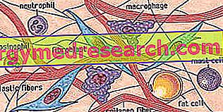
- Collagen fibers They are the most numerous fibers, impart to the tissue in which they are present white color (eg tendons, aponeuroses, organ capsules, meninges, corneas, etc.). They form the scaffolding of many organs and are the most resistant components of their stroma (supporting tissue). They present long and parallel molecules, which are structured in microfibrils, then in long and tortuous bundles held together by a cemented substance containing carbohydrates. These fibers are very resistant to traction undergoing a negligible lengthening. Collagen fibers are composed mainly of a scleroprotein, collagen, a protein far more widespread in the human body, representing 30% of the total proteins. This basic protein is able to change, based on environmental and functional requirements, assuming varying degrees of rigidity, elasticity and resistance. Examples of its range of variability are the integument, the basement membrane, cartilage and bone. - Elastic fibers These yellow fibers predominate in the elastic tissue and therefore in areas of the body where particular elasticity is required (eg ear, skin, pavilion). The presence of elastic fibers in the blood vessels contributes to the efficiency of blood circulation and is a factor that has contributed to the development of vertebrates. The elastic fibers are thinner than the collagen fibers, they branch and anastomose forming an irregular grid, they easily yield to traction forces, resuming their shape when the traction ceases. The main component of these fibers is the elastin scleroprotein, which is somewhat younger, in evolutionary terms, than collagen. - Reticular fibers They are very thin fibers (with a diameter similar to that of collagen fibrils), which can be considered as immature collagen fibers in which they are largely transformed. They are present in large quantities in the embryonic connective tissue and in all parts of the body in which collagen fibers are formed. After birth they are particularly abundant in the scaffolding of the hematopoietic organs (eg spleen, lymph nodes, red bone marrow) and form a network around the cells of the epithelial organs (eg liver, kidney, endocrine glands). |
The connective tissue is morphologically characterized by various types of cells (fibroblasts, macrophages, mast cells, plasma cells, leukocytes, undifferentiated cells, adipose or adipocyte cells, chondrocytes, osteocytes, etc.) immersed in an abundant intercellular material, defined as MEC (extracellular matrix), synthesized by the same connective cells. The ECM is composed of insoluble protein fibers (collagen, elastic and reticular) and fundamental substance, wrongly defined as amorphous, colloidal, formed by soluble complexes of carbohydrates, mostly related to proteins, called acid mucopolysaccharides, glycoproteins, proteoglycans, glucosaminoglycans or GAGs (hyaluronic acid, coindroitinsulphate, keratinsulfate, heparin sulphate, etc.) and, to a lesser extent, from proteins, including fibronectin.
Cells and intercellular matrix characterize various types of connective tissue: connective tissue proper (connective tissue), elastic tissue, reticular tissue, mucous tissue, endothelial tissue, adipose tissue, cartilage tissue, bone tissue, blood and lymph. Connective tissues therefore play different important roles: structural, defensive, trophic and morphogenetic, organizing and influencing the growth and differentiation of surrounding tissues.
Extra Cellular Matrix (MEC)
The conditions of the fibrous part and of the fundamental substance of the connective system are partly determined by genetics, partly by environmental factors (nutrition, exercise, etc.).
Protein fibers are in fact able to change according to environmental and functional requirements. Examples of their structural and functional variability spectrum are the integument, the basement membrane, cartilage, bone, ligaments, tendons, etc.
The fundamental substance continuously changes its state, becoming more or less viscous (from fluid to glue to solid), based on specific organic needs. Can be found in large quantities as synovial joint fluid and ocular vitreous humor, it is actually present in all tissues.
The connective tissue varies its structural characteristics through the piezo-electric effect : any mechanical force that creates structural deformation stretches the inter-molecular bonds producing a slight electrical flow (piezoelectric charge). This charge can be detected by the cells and lead to biochemical changes: for example, in bone, osteoclasts cannot "digest" piezoelectrically charged bone.