Generality
The calcaneus is one of the 7 bones that make up the tarsus of the foot, as well as the bone element constituting the so-called heel.
Protagonist of different articulations - including the ankle joint proper - the calcaneus borders on the talus, superiorly, and with the cuboid bone, anteriorly. Astragalus and cuboid bone are two other tarsal bones.

The calcaneus serves to transfer the body weight to the ground, which weighs on the lower limbs, and to provide the insertion to muscles and ligaments fundamental to plantarflexion, dorsiflexion, eversion and inversion of the foot and flexion movements of the knee.
Brief anatomical reference to the foot
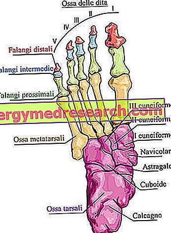
The anatomists divide the bones of the foot into three groups: the tarsal bones, the metatarsal bones and the phalanges.
- Tarsal bones or tarsal group or tarsus . Located just below the ankle joint, there are a total of 7 irregularly shaped bony elements.
- Metatarsal or metatarsal group bones or metatarsals . Belonging to the category of long bones, they are in all 5 elements, arranged parallel to each other. The proximal section is bordered by the cuneiform tarsal bones and the cuboid; the distal section, on the other hand, borders on the phalanges.
- Phalanges . There are a total of 14 and they represent the bony elements that make up the toes. Except the first finger - the only one formed by 2 phalanges - all the other fingers have 3 phalanges each.

What is the heel?
The calcaneus is one of the 7 bones that form the tarsus, as well as the bone that constitutes the anatomical region of the foot called the heel .
Anatomy
Introduction: to fully understand the anatomical references regarding the heel, it is good to remind readers of the names of the other tarsal bones: astragalus, navicular, cuboid, lateral cuneiform, intermediate cuneiform and medial cuneiform.
Known to be the largest tarsal bone, the calcaneus forms the back of the foot as well as the back of the tarsus. Irregular in shape, it resides in close contact with the talus and contributes, along with the latter and the malleoli of the tibia and fibula, to the formation of an important articulation of the human body: the talocrural articulation or ankle properly so called (NB: the only term "ankle" is improper, although it is the one most used).
The heel also has relationships with another tarsus bone: the so-called cuboid bone .
To simplify the anatomical description of a complex bone such as the calcaneus, the experts divide the bony element in question into 6 surfaces (or faces), which are: the anterior surface, the posterior surface, the upper surface, the plantar surface, the lateral surface and the medial surface. As can be easily understood, the adjectives anterior, posterior, superior, plantar, etc. they are used to specify the position of each surface. Then:
- The front surface is the front part;
- The plantar surface is the lower part (plantar refers to the sole of the foot);
- The back surface is the bone portion that lies behind;
- The upper surface is the bone portion above;
- The lateral surface is the bone portion that forms the outer side of the heel;
- The medial surface is the bone portion that forms the inner side of the heel.
In anatomy, medial and lateral are two terms of opposite meaning, which serve to indicate the distance of an anatomical element from the sagittal plane . The sagittal plane is the anteroposterior division of the human body, from which two equal and symmetrical halves are derived.
Mediale means "near" or "closer" to the sagittal plane, while lateral means "far or" farther "from the sagittal plane.
FRONT SURFACE
Of the 6 surfaces in which the calcaneus can be divided, the front surface is the smallest. What makes it important is the presence of an articular surface, through which the calcaneus interacts with the cuboid tarsal bone and gives rise to the calcaneo-cuboid joint .
REAR SURFACE
The back surface has the shape of an irregular dome, with the lower part wider than the upper one.
In the posterior surface, the anatomists recognize three areas, which are: the upper area, the median area and the lower area. Of these three areas, the most important is certainly the median area, as it is the bony portion into which the Achilles tendon is inserted. The precise point of the median area, in which the Achilles tendon is inserted, is a bone growth known as calcaneal tuberosity .
UPPER SURFACE
Anatomically very complex, the upper surface comprises articular portions - that is, with articular function - and non-articular portions .
- There are three joint parts in all; their name is: posterior talar facet, talar mid facet and anterior talar facet. Covered by hyaline cartilage, the posterior, middle and anterior talar facets have the task of articulating the entire calcaneus to the talus, in three different points. The articulation resulting from the relationship between the three facets of the calcaneus and the talus is called the subtalar joint .
- The non-articular portions are varied. Two are reported, namely the posterior portion and the anterior portion.
The first is relevant, because it supports a fat pad, located in front of the terminal tract of the Achilles tendon.
The second is important, because it gives rise to a groove called the heel groove (or calcaneal groove). The groove of the calcaneus is opposed, superiorly, to a very similar area, belonging to the astragalus and called, not surprisingly, the groove of the talus . Together, the groove of the heel and the groove of the talus form the so-called sine of the tarsus . The breast of the tarsus is a small cavity that houses blood vessels, nerves and important ligaments of the foot, such as the interosseous talo-calcaneal ligament and the cervical interosseous ligament. These ligaments are important because they have the function of proprioception during walking and guarantee stability to the foot.

Figure: upper surface of the calcaneus
PLANTAR SURFACE
The plantar surface, or lower surface, has an irregular appearance, with the posterior portion wider than the anterior portion.
It is important because it has two prominences: the so-called calcaneal tubercle, in posterior position, and the so-called anterior tubercle, in the direction of the toes.
- Not to be confused with the calcaneal tubercle of the posterior surface, the calcaneal tubercle of the plantar surface is a rather wide area, which presents at least two areas worthy of mention: the medial process, on the side of the inner edge of the foot, and the lateral process, on the side of the outer edge of the foot.
The medial process is the site of origin of the plantar fascia, of the abductor muscles of the big toe and short flexor of the toes and of one of the two initial ends of the square plantar muscle .
The lateral process, on the other hand, is the point from which the abductor muscle of the fifth toe and one of the two initial ends of the square plantar muscle originate.
- The anterior tubercle is the site of insertion for one of the two ends of the short plantar ligament .

Figure: plantar surface of the calcaneus
What is the plantar fascia?
The plantar fascia, or plantar aponeurosis, is a sort of very thick ligament, located on the lower edge of the foot (sole of the foot), which runs from the plantar surface of the calcaneus to the bones of the fingers. Morphologically similar to an arch, it allows the curvature of the foot and acts as a cushion that absorbs the shocks of walking, running, etc.
The plantar fascia is known to most people because it is the protagonist of the medical condition known as plantar fasciitis (see chapter on pathologies).
SIDE SURFACE
Rough and flat, the side surface is wider at the back than at the front.
Anteriorly, it presents a small excrescence, called the fibular trochlea .
Almost in the middle, on the other hand, it has a small prominence, on which a head of the calcaneo-fibular ligament is inserted.
On the lateral surface, there are also two grooves, one located higher than the other. The upper groove is the site of passage of the terminal tendon of the short peroneal muscle, while the lower groove is the site of passage of the terminal tendon of the long peroneal muscle .

Figure: lateral surface of the heel
MEDIUM SURFACE
The medial surface is an area with a hollow appearance, presenting a bony projection that the anatomists have called such sustentaculum . Called this way because it supports the medial portion of the talus, the sustentaculum has two anatomically relevant areas: the lower surface and the medial margin.
The lower surface is important, because the tendon of the long flexor muscle passes over it.
The medial margin, on the other hand, is important, because it gives insertion to one of the two ends of the plantar calcaneo-navicular, tibio-calcaneal and medial astragalocellar ligaments .
JOINTS
The heel participates in four joints :
- The aforementioned talocrural joints, calcaneo-cuboid and subthalar. Remember that the first involves heel, astragalus and malleolus; the second is the result of the synergism between calcaneus and cuboid bone; finally, the third is the result of the interaction between calcaneus and astragalus.
- The calcan-navicular joint, whose ratio between the heel and navicular bone depends on the presence of some ligaments.
Participation in the talocrural and subtalar joints occurs through the upper portion of the calcaneus, while participation in the calcaneus-cuboid and calcaneus-navicular joints occurs through the anterior portion of the calcaneus.
LIGAMENTS
In summary, the ligaments that have a relationship with the heel are:
- The calcareous -navicular plantar ligament, the tibio-calcaneal ligament and the medial talar-calcaneal ligament, at the level of the medial surface of the calcaneus.
- The calcaneo-fibular ligament, at the level of the lateral surface of the calcaneus.
- The plantar ligament short, at the level of the plantar surface of the calcaneus (NB: the planar fascia, present on the plantar surface, is not normally considered a true ligament).
- The interosseous talo-calcaneal ligament and the cervical interosseous ligament, at the level of the upper surface of the calcaneus.
OSSIFICATION
The ossification process of the heel has two centers of ossification as protagonists: a primary center and a secondary center.
The primary ossification center begins its activity in the third month of an individual's intrauterine life. The secondary ossification center, on the other hand, is activated between the 6th and 8th year; it resides on the back surface.
The fusion of the bone portions, generated by the two centers, takes place around the age of 14-16.
vascularization
The flow of oxygen-rich blood to the calcaneus depends on the branches of the posterior tibial artery and the perforating arteries, which derive from the peroneal (or peroneal) artery .
Function
The calcaneus covers various functions.
First, it contributes, along with the other bones of the foot, to the transfer to the ground of all the body weight, which is serious on the lower limbs.
Secondly, it offers a fundamental contribution to locomotion and to the possibility of performing motor activities such as running or jumping, as on it the initial or terminal heads of different muscles and some ligaments of the ankle are inserted.
The table below summarizes all the muscles that relate to the heel, specifying the bone surface with which the muscles in question come into contact.
| Heel surface | Muscles | Initial boss or terminal boss | Contact site on the heel |
| Back surface | Gastrocnemius muscle Soleus muscle Plantar muscle | Head end Head end Head end | For all three the insertion takes place on the calcaneal tuberosity, through the Achilles tendon. |
| Plantar surface | Big-tooth abductor muscle Short flexor muscle of the fingers Fifth finger abductor muscle Square plantar muscle | Initial leader Initial leader Initial leader Initial leader | Media process. Media process. Lateral process. Medial and lateral processes. |
| Back surface | Short extensor muscle of the fingers Short extensor muscle of the big toe | Initial leader Initial leader | For both, the place of origin lacks a precise name. |
* NB: some texts on human anatomy report that one of the two initial ends of the square plantar muscle originates from the medial surface.
CALCANNO AND MOVEMENT CAPACITY
Plantarflexion, dorsiflexion, eversion and inversion of the foot, and knee flexion movements depend on the muscles and ligaments of the ankle that are related to the heel.
Furthermore, the same muscles and the same ligaments of the ankle play a fundamental role in stabilizing the ankle leg, while the human being is in an upright position.
At this point, it is worth remembering that:
- The plantarflexion of the foot is the movement that allows you to point your foot towards the floor. The human being performs a plantarflexion movement when he tries to walk on his toes.
- Dorsiflexion is the movement that allows you to lift your foot and walk on your heels.
- Eversion means raising the side edge (ie the outer edge) of the foot, keeping the medial edge (ie the inner edge) on the floor.
- Inversion means raising the medial edge of the foot, keeping the side edge on the floor.
- The knee flexion is the movement that allows you to bend the knee, so as to reduce the angle between the leg and the thigh.
Associated pathologies
Like all bony elements present in the human body, the calcaneus can also fracture.
Typically, fractures of the calcaneus are the consequence of impacts that affect the heel and violently push the heel against the talus.
The main circumstances that cause a heel fracture are falls on the heels, especially those from great heights.
From the point of view of their diffusion in the general population, calcaneus fractures represent 2% of all fractures, which can affect the human body, and 60% of all fractures, which can affect the tarsal foot.
The typical symptoms of a fracture of the heel consist of: foot pain, walking instability, difficulty in performing movements with the affected foot, presence of swelling, presence of hematoma and redness.
The fractures of the calcaneus are injuries that can give rise to various late complications, on all the arthritis against the subtalar joint and the strong pain during the movements of eversion and inversion of the foot.
The treatment of a calcaneus fracture can be conservative or surgical.
In general, the conservative approach is reserved for less severe calcaneus fractures, while the surgical approach is reserved for more severe calcaneus fractures.