Generality
The chest CT scan is the Computerized Axial Tomography that allows to study in detail, and from multiple angles, the state of health of the anatomical structures of the thoracic area of the human body (ie lungs, heart, sternum, ribs, thoracic tract of the spine, blood vessels, esophagus, etc.).

Thanks to a chest CT scan, the radiologist is able to identify: lung tumors, chronic respiratory diseases, the exact reason of certain symptoms, lesions of the thoracic organs or blood vessels, pulmonary metastases, the characteristics of blood flow in the arteries and veins of the thorax, etc.
The main risks of chest CT are related to the dose of ionizing radiation, to which the patient is exposed during the examination, and - if used - to the contrast agent (which in some individuals is the cause of an allergic reaction).
Contraindicated in case of pregnancy, the chest CT provides excellent quality images.
Short review of what the TAC is
TAC, or Computerized Axial Tomography, is a diagnostic procedure that uses ionizing radiation ( X-rays ) to create extremely detailed three-dimensional images of specific anatomical areas of the body (brain, bones, blood vessels, abdominal organs, thoracic organs, pathways respiratory, etc.).
The TAC equipment includes:
- The large donut-shaped scan unit, called a gantry . It is the radioactive source;
- The generator;
- The support on which to place the patient. Generally, it is a sliding bed;
- An electronic processor;
- A command console for displaying three-dimensional images;
- A system for recording the acquired data.
In addition to the conventional CT (or classical or without contrast ), there is also the CT with contrast medium : the contrast medium allows to obtain images richer in details of a particular organ or anatomical tissue and of the blood vessels.
The TAC is normally painless ; it can be slightly painful, when intravenous injection of the contrast medium is expected.
Nevertheless, it is in any case among the minimally invasive procedures, since the dose of ionizing radiation to which the patient is exposed is considerable.
What is the chest CT scan?
Thoracic CT, or chest CT, is the diagnostic test that exploits the potential of Computerized Axial Tomography to visualize, in detail and from multiple angles, the various anatomical structures of the thorax (heart, lungs, mediastinum, large vessels such as the aorta, the pulmonary artery and the vena cava, the bones of the thoracic cage, the esophagus etc.).
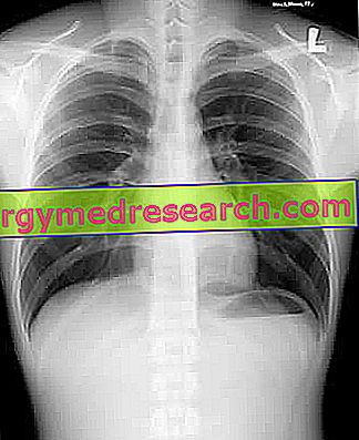
Definitely more precise than the so - called RX-thorax (the classic chest radiograph ), the chest CT scan is a radiology procedure, exactly like all other types of CT scans.
The interpretation of the images of the chest CT scan is up to a radiologist, who, if necessary, will advise the patient to discuss what has emerged with a specialist of the respiratory system and / or the cardiovascular system (a pulmonologist or a cardiologist).
TAC thorax with contrast medium
Like many other types of CT scans, chest CT is also practicable in " contrast " or " contrast " mode.
In general, the contrast media used are iodine- based ( iodinated contrast media ).
Indications
The chest CT allows:
- To investigate the findings of an abnormal chest X-ray;
- Understanding the exact origin of symptoms, such as coughing, chest pain, dyspnoea, etc., which lead to thinking about the presence of a heart disease or a respiratory illness;
- Detect lung tumors and interpret their severity;
- Determine lung metastases;
- Evaluate whether the tumors that can affect the thoracic organs (NB: lungs in most cases) are responding or not to treatments;
- Plan a radiotherapy treatment;
- Estimate any damage to the thoracic organs, heart and lungs, to the large blood vessels of the chest, to the ribs, to the sternum and / or to the vertebral tract of the spine.
Lung diseases identifiable by chest CT scan
The chest CT scan is one of the diagnostic tests of choice for the detection of lung diseases .
Among the possible pulmonary diseases identifiable by means of chest CT, we note in particular:
- Benign and malignant lung tumors;
- Pneumonia;
- Tuberculosis;
- Cystic fibrosis;
- Bronchiectasis;
- Pleural diseases (pleurisy, pleural mesothelioma, pleural effusion, pleural empyema, etc.);
- Chronic diseases of the lungs (eg: pulmonary fibrosis, chronic obstructive pulmonary disease, etc.) or pulmonary interstitium (eg, interstitial disease);
- Congenital anomalies of the lungs and large blood vessels that, with origin in the heart, reach the lungs.

Indications for chest CT with contrast medium
Generally, physicians resort to chest CT with contrast medium, when they need to assess blood flow within the arterial and venous vessels of the chest.
Preparation
The chest CT without contrast medium does not require special preparation. In fact, the only preparatory rules that the patient must follow are:
- Tell the doctor who prescribes the exam if you suffer from claustrophobia . In such cases, the solution is the use of a sedative;
- In the event that the patient is a woman, communicate any pregnancy or presumed status;
- Take the exam without jewelry or clothing with metal parts, as these could interfere with the proper functioning of the diagnostic equipment.
If the patient does not comply with this preoperative rule, he is invited to do so just before the procedure begins.
Preparation in case of chest CT with contrast medium
If the chest CT scan involves the use of the contrast agent, the exam preparation is more articulated than previously stated. In fact, besides providing the three previous recommendations, the patient must:
- Communicate to the doctor who prescribes the exam if you suffer from any allergy, in particular to the iodine in contrast media;
- Tell your doctor if, at that time in your life, you are taking medication, both against temporary conditions and against chronic conditions;
- Inform your doctor of any illness suffered in the last period and if you suffer from a heart disease, diabetes, asthma, a thyroid disease and / or a kidney disease ;
- Introduce yourself to fast food for at least 6-8 hours. This means that, if the chest CT is held in the morning of a certain day, the last meal must be the dinner of the previous evening.
Procedure
First, at the invitation of a medical staff member, the patient must:
- Answer a quick questionnaire about your medical history,
- Wear a special coat instead of your clothes and finally
- Take off all the jewelery and other metal objects that it is wearing, for the reasons stated above.
Once all these preliminary operations have been completed, the patient is ready to take his seat on the sliding table of the chest CT, which serves, at a later time, to position him inside the so-called gantry (see figure).

To lie correctly on the couch, the patient can count on the help of a member of the medical staff (in general it is the same person who previously questioned him about his medical history, measured his pressure and temperature, etc.); during his placement, the one who helps him also reminds him how he must "behave" during the examination and the importance that his immobility has, when the instrument is in operation.
Once the patient is lying on the couch with all the necessary comforts (pillows, blankets, earplugs, etc.), the introduction of the sliding couch in the gantry can finally take place and the phase dedicated to the creation of three-dimensional images begins.
The creation of the images is very noisy: this explains the supply to the patient of earplugs or, alternatively, of a pair of headphones.
It is good to remember that, while the diagnostic device is in operation, the entire medical staff leaves the room where the patient under examination resides and moves to an adjacent room, in which there are the command console of the gantry and the system of registration of the acquired data. However, the patient is not really alone: in fact, the room in which he finds himself is equipped with a loudspeaker and a camera, through which he can communicate with the outside in case of sudden necessity.
After collecting the images necessary for a detailed evaluation of the thoracic compartment, the radiologist declares that the chest CT scan is complete and starts the operations to extract the patient from the gantry, an extraction in which the "usual" medical staff member is already involved. times, in the previous phases.
Getting up from the couch and getting dressed, the patient is ready to go home and to his daily activities, unless otherwise indicated by the radiologist.
Important note on immobility during the examination: in some stages of chest CT, the patient is also invited to hold his breath
How does the chest CT with contrast medium vary in the procedure?
In addition to what was previously reported, the chest CT with contrast includes the injection of the latter. This fundamental step takes place shortly after the patient is settled, but a few minutes before the bed is placed in the gantry .
Injecting the contracting medium is the radiologist, in collaboration with a registered nurse.
The injection site is a vein in the arm or hand; the achievement of the anatomical areas of diagnostic interest, by the contrast medium, takes a few minutes.
What feelings does the patient feel during the procedure?
Without the use of contrast medium, the chest CT scan is a painless procedure, which takes place in a fairly linear manner (ie it does not induce particular sensations).
With contrast media, on the other hand, it could cause discomfort, at the time of inserting the needle for injection of the contrast medium, and a strange metallic taste in the mouth, starting from a couple of minutes after the injection of the aforementioned contrast agent.
How long does the chest CT scan last?
Generally, chest CT lasts a maximum of 20-30 minutes .
The possible use of the contrast medium increases the aforementioned time by a few minutes.
On what occasions can the patient not return home immediately?
If the chest CT scan provides unclear images, the patient is forced, on the decision of the radiologist, to postpone the return home, to repeat the entire procedure.
How to encourage the elimination of contrast media?
To facilitate the elimination of the contrast agent from the body (clearly in cases of contrasted chest CT), radiologist doctors indicate that they drink a lot of water .
If the patient adheres to this indication, he eliminates the contrast agent from his body within 24 hours of completing the chest CT scan.
risks
With or without contrast, the chest CT scan involves exposing the patient to a certain dose of ionizing radiation, which - as is well known - represent a factor favoring the onset of malignant neoplasms .
With regard to this topic, we take this opportunity to inform readers that, thanks to advances in medical technology in the field of radiology, today there are apparatuses for thoracic CT scans with low emission of ionizing radiation .
Certainly less dangerous than standard instruments, these modern chest CT scans, while using a lower dose of ionizing radiation, guarantee good quality images, useful for the diagnosis of many pulmonary and cardiovascular problems.
In any case, apart from the doses of ionizing radiation emitted, the radioactive risk connected to the chest CT imposes a certain balance in prescribing the repeated execution of this diagnostic test.
Radioactivity level of a low-dose CT scan
A "standard chest CT scan" exposes patients to a dose of ionizing radiation equivalent to 2 years of natural radioactivity; a "low-dose chest CT scan", on the other hand, exposes patients to a dose of ionizing radiation equivalent to only 6 months of natural radioactivity (so it is 4 times lower than in the previous case).
Risks of chest CT with contrast medium
In some predisposed individuals, the use of contrast agents may give rise to allergic reactions, the most common symptoms of which are: hot flushes, nausea, strange tingling, hives and prolonged pain at the injection site.
Allergic reactions to contrast agents for chest CT scans are rare circumstances which, with the necessary drugs, are both controllable and preventable.
Contraindications
In the case of chest CT without contrast medium, the only contraindications are: the state of pregnancy and obesity (the CT scans can generally support people weighing no more than 150 kilograms).
In the case of chest CT with contrast medium, however, there are further contraindications: in fact, the aforementioned pregnancy and obesity are compounded by the presence of an allergy or an intolerance towards the contrast agent and renal insufficiency (the whose presence prevents the correct elimination of the contrast agent).
Is breastfeeding a contraindication to the use of contrast media?
Breastfeeding is not a contraindication to chest CT with contrast medium; however, for most doctors and those who produce contrast media, it is not recommended in the first 24-48 hours following the exam.
Results
Depending on the severity of the patient's condition, chest CT results may be available immediately or after a few days.
The main advantage of the chest CT scan is that it provides very valuable information for a correct diagnosis.
Other advantages of chest CT:
- It is painless, minimally invasive and very precise;
- Simultaneously shows the so-called soft tissues (nerves, muscles, ligaments, fat, blood vessels etc.) and the so-called hard tissues (bones and cartilages);
- Provides more detailed images of conventional X-rays;
- It is a rapid examination, of short duration;
- Identifies many morbid conditions;
- It is less sensitive to patient movements than nuclear magnetic resonance;
- Unlike nuclear magnetic resonance, it can be performed even if the patient has a metal prosthesis;
- It provides images in real time, so surgeons can take advantage of it just before surgery;
- Thanks to its high detection capacity, it could make the use of exploratory surgery or a biopsy superfluous.


