Generality
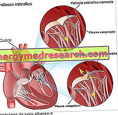
The mitral valve prolapse ( PVM ), or mitral valve prolapse, consists of a wrong movement, at the time of closure, of the flaps (or cusps) that constitute the mitral valve of the heart.
Placed in control of the blood flow between the left atrium and the ventricle, the mitral valve, if not correctly closed at the time of the systole, causes a blood regurgitation towards the left ventricle → left atrium. For this reason, mitral valve prolapse is one of the causes of mitral insufficiency. The abnormal positioning of the cusps is caused by a degeneration of the cuspid tissue itself or by the rupture of one of the structural elements of the mitral valve.
What is the mitral valve prolapse
The mitral valve prolapse ( PVM ), or mitral valve prolapse, consists of an abnormal closing movement of the cusps (or flaps) that constitute the mitral (or mitral) valve of the heart. Under normal conditions, the mitral valve controls blood flow in the left atrium direction - left ventricle and prevents reflux in the opposite direction during ventricular systolic due to hermetic closure. At the onset of a mitral valve prolapse, instead, during the contraction phase of the ventricle (ventricular systole), a share of blood, instead of turning into the aorta, goes back and goes back to the left atrium; this occurs because the valve orifice is not completely closed. This is the so-called regurgitation of blood, which characterizes another important heart disease: mitral insufficiency ; we will see later that the two valve defects, prolapse and mitral insufficiency, are closely related.
Mitral valve prolapse affects women more than men. It is also more frequent in long-limbed subjects, with elongated and flattened thorax, as well as individuals suffering from dorsal scoliosis.
Before proceeding with the description of the main causes that determine a mitral valve prolapse, it is good to recall some fundamental characteristics of the mitral valve. Recalls that will also be useful to describe the appearance and functioning of the same valve when it is subject to prolapse, that is, respectively, the pathological anatomy and the pathophysiology.
Therefore:
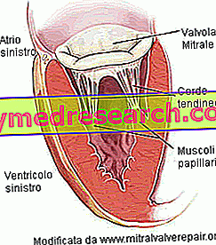
- The valve ring . Circumferential structure of connective tissue defining the valve orifice.
- The valve orifice measures 30 mm in diameter and has a surface of 4 cm2.
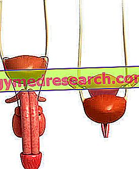
- Two flaps, front and back. It is said, for this reason, that the mitral valve is bicuspid . Both flaps fit into the valve ring and look towards the ventricular cavity. The anterior leaflet looks towards the aortic orifice; the posterior flap faces, instead, on the wall of the left ventricle. The flaps are composed of connective tissue, rich in elastic fibers and collagen. To facilitate the closure of the orifice, the edges of the flaps have particular anatomical structures, called commissures. There are no direct controls, of the nervous or muscular type, on the flaps. Similarly, there is no vascularization.
- The papillary muscles . They are two and are extensions of the ventricular musculature. They are sprayed by the coronary arteries and give stability to the tendinous cords.

Causes of mitral valve prolapse.
Pathological anatomy and pathophysiology
The main cause of mitral valve prolapse is the degeneration of the loose connective tissue that constitutes the flaps (or cusps) of the mitral valve. It is a myxomatous degeneration, as the middle layer of the connective tissue of the valve leaflets is subject to myxoma . The myxoma is a particular neoplastic form (tumor), at which the extracellular matrix that makes up the cardiac connective tissues is altered; therefore, the matrix varies in composition and we have that:
- Collagen fibers are inadequately produced.
- The mucopolysaccharides of the basic substance increase in quantity.
Myxomatous degeneration occurs in some components of the mitral valve and changes its morphology:
- The valve leaflets become more elongated, yielding and thickened.
- The tendinous cords stretch and can sometimes even break.
- The valve ring increases its circumference.
Altered in the structure, the cusps no longer seal the valve orifice in a hermetic way.
Failure to close the valve is usually due to only one of the flaps, the rear one. Sometimes, however, both are interested. The anomaly, in the closing movement, consists in a flexion of the flaps towards the atrial cavity. In other words, if in normal conditions, the flaps turn towards the ventricle, in cases of prolapse, they curve from the opposite side, towards the cavity of the left atrium. In fact, the term prolapse means the outflow of a bowel from the cavity in which it is contained through a natural opening. The definition is reminiscent of a hernia. In the specific case, we are not talking about a real hernia, since the bowel in question is a flap of the valve, but the behavior is very similar.
The alteration of the normal mitral valve closure, during systole, causes the same physiopathological adaptations that characterize mitral insufficiency. Then:
- The regurgitation of blood flows into the left atrium and enlarges its size. Cardiac output is depleted due to regurgitation. Therefore, blood circulation is inefficient. The individual faces this situation by increasing the breaths.
- At the next diastole, the mitral valve opens, causing the regurgitation to flow from the atrium to the left ventricle. This is a situation that normally does not occur and has consequences for the pressure gradient between atrium and ventricle.
- Regurgitation, inside the ventricle, raises the ventricular pressure, altering the normal balance with the atrial pressure value. A situation called left ventricular failure is determined.
These three repercussions on blood flow are not always equally critical. In other words, mild forms of mitral valve prolapse result in mild mitral insufficiency. The same can be said of moderate forms, while the case in which another cardiac disease is associated with mitral prolapse is quite different: the consequences on blood flow are more serious.
Although less frequent, there are other causes that cause mitral valve prolapse.
- Marfan syndrome
- Ehlers-Danlos syndrome
- Rheumatic endocarditis
- Ischemic heart disease
- traumas
- Obstructive hypertrophic myocardiopathy
- Surgery operations on the mitral valve
- Lupus erythematosus
- Duchenne muscular dystrophy
- Interatrial septal defect
- Hyperthyroidism
- Turner syndrome
- Ebstein's disease
These include Marfan syndrome and Ehlers-Danlos syndrome . They are two congenital pathologies, that is, present since birth. They determine alterations in the connective tissues that follow the structural and morphological changes, induced by myxomatous degeneration, described above.
Symptoms and signs
Mitral valve prolapse shows a symptomatology very similar to that of mitral insufficiency. However, it is fair to point out that, in most cases, mitral prolapse is asymptomatic, that is, it has no symptoms. In this case, the individual carrying this anomaly has a normal life, can play sports and perform any other physical activity of a healthy person.
The most frequent symptoms are:
- palpitations
- Dyspnea on exertion
- Asthenia
- Chest pain
- Vertigo
- Syncope
Heart disease, also known as palpitation, is the most frequent symptom in those who experience a mitral valve prolapse. Heart failure is an increase in the intensity and frequency of the heartbeat; it usually manifests itself with a tachycardia, that is an increase in the speed of the heartbeat, but it can sometimes give rise to different types of arrhythmias . Arrhythmias are changes in the normal heart rhythm. Heart rhythm that originates from a natural pacemaker, known as an atrial sinus node . Ventricular extrasystoles and atrial fibrillation are reported among moderate and severe arrhythmias, respectively.
Ventricular extrasystoles consist of a contraction of the heart that occurs earlier than the regular heart rhythm, altering the succession of beats. It can be an isolated or repeated phenomenon: if repeated, the extrasystole is much more dangerous. Furthermore, the isolated extrasystole is much more frequent, in terms of appearance, compared to repeated extrasystoles and atrial fibrillation.
Atrial fibrillation is a cardiac arrhythmia, ie an alteration of the normal heartbeat rhythm. It is due to a disorder of the nerve impulse coming from the atrial sinus node. It results in fragmentary and ineffective atrial contractions from a haemodynamic point of view (ie what concerns the blood flow). In the case of a mitral valve prolapse, the regurgitation of blood in the atrium reduces the blood volume pushed into the aorta by ventricular contraction. In light of this, the body's oxygen demands are no longer satisfied. Faced with this situation, the individual suffering from atrial fibrillation increases the respiratory acts, manifest palpitation, irregularity of the wrist and, in some cases, fainting due to lack of air. The picture can further degenerate: an ever increasing regurgitation and the accumulation of blood in the vascular systems upstream of the left atrium, if associated with an altered coagulation, give rise to the formation of thrombi (solid, non-mobile masses, composed of platelets) inside the vessels. The thrombi can disintegrate and release particles, called emboli, which, traveling in the vessel system, can reach the brain, or the heart. In these locations, they become an obstacle to normal spraying and oxygenation of brain or cardiac tissues, causing the so-called ischemic stroke (cerebral or cardiac) situation. In the case of the heart, one also speaks of a heart attack . In subjects suffering from mitral valve prolapse, it is however a rare event.
Effortless dyspnea consists of difficult breathing. In the specific case, it results from the decreased cardiac output of the left ventricle, due to the proportion of blood regurgitated towards the left atrium. Therefore, the organism's response is to increase the number of breaths, in order to offset the volume of the range.
Similarly, syncope is another natural consequence of the compromised efflux of blood from the left ventricle and directed to the brain. In fact, syncope occurs when blood flow to the brain tissue is reduced. The lower cardiac output, associated with mitral prolapse, prevents normal blood supply to the brain tissue and this condition can manifest itself either during an effort, or physical activity, or, serious eventuality, at rest. Syncope at rest is often associated with a malfunction of the left ventricle and can cause sudden death. In those who suffer from mitral valve prolapse, this is a rare event; the feeling of vertigo is instead much more common, also linked to the lower oxygenation of the brain.
Chest pain, due to angina pectoris, is a rare event. Angina pectoris, in this case, is due to left ventricular hypertrophy, ie the left ventricle, and not to an occlusion of the coronary vessels. In fact, the hypertrophic myocardium needs more oxygen, but this request is not adequately supported by the coronary implant, which remains unchanged. Therefore, an imbalance occurs between the consumption and the supply of oxygen to the tissues. The typical pain of angina pectoris is felt in the left hemithorax.
Asthenia is a sense of weakness and lack of energy.
The characteristic clinical signs of a prolapse of the mitral valve are two:
- The click . It is a noise caused by modified tendon strings.
- The systolic murmur . It originates from the regurgitation of blood, through the defective valve, during ventricular systolic contraction.
Both are revealed through listening.
Diagnosis
Mitral insufficiency can be detected by the following diagnostic tests:
- Stethoscopy.
- Electrocardiogram (ECG).
- Echocardiography.
Stetoscopy . Detection of a systolic murmur is one of the most important clues for diagnosing a mitral valve prolapse. The sound of the breath is produced by the passage, from the left ventricle to the left atrium, of the regurgitation of blood. It is perceived in the systolic phase, since it is at this moment that the mitral valve is not closed as it should. The detection zone is in the V intercostal space, ie the one coinciding with the position of the mitral valve. The other important diagnostic sign, the click, varies in intensity based on the positions assumed by the individual who presents it.
ECG . By measuring the electrical activity of a heart with mitral valve prolapse, the ECG shows the great variety of arrhythmias that can occur in a patient. The list is drawn up on the basis of frequency and hazard characteristics: it starts from the most frequent and least dangerous; it ends with the least frequent but most dangerous.
- Isolated ventricular extrasystoles.
- Tachycardia.
- Atrial fibrillation.
- Repeated ventricular extrasystoles
Diagnosis by ECG gives an idea of the degree of severity of the mitral valve prolapse: if the outcome is comparable to that of a healthy individual, it means that it is not a severe form; vice versa, the examination shows the mentioned irregularities.
Echocardiography . Taking advantage of the ultrasound emission, this diagnostic tool shows, in a non-invasive way, the fundamental elements of the heart: atriums, ventricles, valves and surrounding structures. From echocardiography, the doctor can detect:
- Abnormal behavior of the flaps and tendon strings of the valve.
- Anomalies of the left ventricle, during the phases of systole and diastole.
- Increase in the size of the left atrium (dilated atrium).
- The maximum flow velocity is the turbulent systolic flow of the regurgitation, employing continuous and pulsed Doppler techniques, respectively. From the first measurement, the pressure gradient between the left atrium and the left ventricle can be derived; from the second, the extent of regurgitation.
Therapy
The medical treatment of mitral valve prolapse, from the less severe and asymptomatic to the severe cases, is very similar to that of mitral insufficiency. The therapeutic approach therefore varies according to the severity of the heart disease. The asymptomatic forms, but also the mild ones, require preventive measures, designed to avoid bacterial infections, such as endocarditis, which affect the cardiac cavities. Periodic check-ups are also recommended every 2-3 years, but the individual, who carries a mild form of prolapse, can perform any activity, including sports. The most used drugs, in the mild forms of mitral valve prolapse, are:
- Beta-blockers and anxiolytics . They are used when mild arrhythmias occur.
The first appearance of symptoms and moderate / severe forms require more attention: in addition to drug therapy, surgery can become decisive.
The critical situations, which advise the intervention, are:
- The ascertained rupture of the tendon cords of the valve.
- Repeated and gradually more serious arrhythmias.
- Ascertained increase in atrial cavity, following regurgitation
- Left ventricular decompensation.
These clinical findings are similar to those that occur during moderate / severe chronic mitral insufficiency.
There are two possible surgical operations:
- Replacement of the valve with a prosthesis . It is the most implemented intervention for the valves of those individuals, not young, with serious anatomical anomalies. A thoracotomy is performed and the patient is placed in extracorporeal circulation (CEC). Extracorporeal circulation is achieved through a biomedical device which consists of creating a cardio-pulmonary pathway that replaces the natural one. In this way, the patient is guaranteed an artificial and temporary blood circulation that allows surgeons to interrupt the flow of blood in the heart, diverting it to another equally effective path; at the same time, it allows you to operate freely on the valve apparatus. Prostheses can be mechanical or biological. Mechanical prostheses require, in parallel, anticoagulant drug therapy. Biological prostheses last 10-15 years.
- Mitral valve repair . It is the most suitable approach for mitral insufficiencies of "non-rheumatic" origin. In other words, those caused by a mitral valve prolapse. The valvular structures of the ring, of the cusps and / or of the tendinous cords are compromised. The surgeon acts differently, depending on where the valve lesion resides. Again, patients are placed in extracorporeal circulation. It is an advantageous technique, as the prostheses have some drawbacks: the biological ones must be replaced after about 10-15 years; the mechanical ones require the continuous administration, in parallel, of anticoagulants.