Generality
The urethra is the conduit which, starting at the level of the bladder and ending at the level of the urinary meatus, serves mainly to expel urine.
From the histological point of view, it presents various epithelia - including the so-called urothelium (or transitional epithelium) - and two cassocks: the mucous membrane and the muscular cassock.
The most well-known and widespread pathological conditions that can affect the urethra are urethral stenosis - which is the narrowing of the urethra - and urethritis - which is the inflammation, often on an infectious basis, of the urethra.
Brief anatomical reference of the urinary tract
The elements that make up the urinary tract are the kidneys and the urinary tract .
The kidneys are the main organs of the excretory system. In number of two, they reside in the abdominal cavity, on the sides of the last thoracic vertebrae and of the first lumbar vertebrae, they are symmetrical and possess a shape that resembles that of a bean.
The urinary tract, instead, form the so-called urinary tract and have the following structures:
- The ureters . In number of two, it is the ducts that connect the kidneys to the bladder. For the avoidance of doubt, it is specified that each ureter is independent of the other.
- The bladder . It is a small hollow muscular organ, which accumulates urine before urination.
- The urethra . In the aforementioned article, the reader will find all the useful information relating to the anatomy and function of the urethra.

What is the urethra?
The urethra is the channel, of tubular form, which connects the bladder to the so-called urinary meatus (or external urethral orifice ) and which is used for the expulsion from the body of some body fluids (mainly urine).
In anatomy, the term meatus indicates an orifice that connects the inside of the body to the outside and through which, in some cases, liquid passes.
In the human body, there are numerous meatus: the urethral meatus, which is the orifice through which each ureter opens into the bladder; the external acoustic meatus, which is the discrete-sized hole between the pavilion and the tympanic membrane; and so on.
Anatomy
Except for the point of origin (the bladder), the male urethra has some substantial differences from the female urethra. Therefore, they will be treated distinctly, so as to make the description of this important element of the urinary tract clearer.
MALE URETRA
In humans, the urethra is about 15-20 centimeters long, it first crosses the prostate and then the penis (the male reproductive organ) and ends at the tip of the glans (which is the distal end of the penis).
To simplify the study of the urethra, the anatomy experts identify 4 sections (or parts), placed in succession between them:
- The pre-prostatic (or intramural ) section. The urethra is born inside the bladder, in a specific region that takes the name of the neck of the bladder and in which resides the so-called internal urethral orifice .
The pre-prostatic section is the part of the urethra between the bladder neck and the internal urethral sphincter . Its length varies from 0.5 centimeters in some individuals to 1.5 centimeters in others.
To mark the boundary between the pre-prostatic section and the subsequent prostatic section is where the urethra begins crossing the prostate.
- The prostatic section . It is the part of urethra that passes through the prostate. Thanks to this crossing, the prostate communicates with the urethra and introduces (when required) the fluids necessary for reproductive activity (seminal fluid, semen, etc.).
Two different types of channels guarantee the passage of reproductive fluids from the prostate to the urethra: the ejaculatory ducts and the prostatic ducts .
The ejaculatory ducts introduce the spermatozoa into the urethral duct, which come from the vas deferens of the testicles, and the fluid produced by the seminal vesicles.
The prostatic ducts, on the other hand, pour the actual seminal fluid into the urethra, which, by mixing with the spermatozoa and the seminal vesicle fluid, forms sperm .
- The membranous section . It is the tract of urethra that resides between the so-called pelvic floor and the so-called deep perineal space.
1-2 centimeters long and particularly narrow, the membranous section passes through the external urethral sphincter .
At this point, it is worth remembering that the external urethral sphincter and the aforementioned internal urethral sphincter are both two muscular structures, which control the emission of urine; however, while the former (outside) is voluntary, the latter (the inside) is involuntary.
- The spongy section . End section of the urethra, is the section that crosses the entire spongiosa body of the penis and ends at the glans (with the urinary meatus).
The spongious body of the penis is a cylindrical structure of erectile tissue, which resides in the center of the ventral side of the male reproductive organ. Above him, there are two very similar elements, in shape and histology, which are called cavernous bodies.
Generally 15-16 centimeters long, the spongy section has two important peculiarities.
The first peculiarity is that, at the level of the glans, the urethra widens in an evident manner, giving rise to a dilatation that takes the name of navicular fossa of the urethra .
The second peculiarity is that, on this stretch of urethra, the urethral glands and the openings of the two bulbourethral glands take place.
The urethral glands (or Littre's glands ) produce a mucoid substance (mucus), rich in glycosaminoglycans, which serves to protect the internal epithelium of the urethra from the corrosive substances contained in the urine.
The openings of the two bulbourethral glands, on the other hand, serve to enter, in the terminal part of the urethra, an essential substance of the ejaculate and with lubricating function of the urethral duct itself. The secretion of the bulbourethral glands is light in color and contains, mainly, mucoproteins.
Length of the male urethra, during the various stages of life
For obvious reasons, the male urethra stretches along with individual body growth.
Generally, in newborns, it has an average length of about 6 centimeters; in puberty, it is no more than 12 centimeters long; at the end of puberty, it is developed almost definitively.

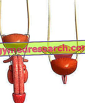
Figure: male urethra.
FEMALE URETRA
The female urethra is 4 centimeters long on average, much shorter than the male urethra.
His path to the urinary meatus begins at the neck of the bladder (where the so-called internal urethral orifice resides), passes through the so-called urogenital trigone (or urogenital diaphragm ) and, resting on the anterior wall of the vagina, opens into the upper part of the vestibule of this (with the "usual" external urethral orifice)
Some anatomy texts report that the position of the urinary meatus is between vagina and clitoris, at 29 millimeters from the latter (NB: the clitoris resides anteriorly to the vagina).
Beyond the shorter length (which, of course, also involves a different path), the female urethra is distinguished from the male by the position of the urethral sphincters. In fact, if in man the internal urethral sphincter localizes before the prostate and the external one after the prostate, in the woman the two aforementioned sphincters are arranged almost in succession, therefore very close to each other.
Sections of the female urethra
The anatomists recognize three segments (or sections) in the female urethra: the intramural (or intravesical) segment, the free segment and the vaginal segment.
The intramural segment runs from the internal urethral orifice to the internal urethral sphincter (as in humans).
The free segment is the tract that begins after the internal urethral sphincter, passes through the urogenital trigone and ends before entering into close relationship with the vagina.
Finally, the vaginal segment is the section intimately adherent to the vagina and ending with the external urethral orifice.

Figure: female urethra
TONACHE AND EPITHELI OF THE URETHRA: A LITTLE HISTOLOGY
Between cassocks (that is membranes) and epithelia, the male urethra and the female urethra have a rather particular structure, worthy of a brief but significant description.
Epithelia . The first features of the male urethra and of the female urethra present a transitional epithelium. This epithelium is typical of the urinary tract, so that experts also call it urothelium.
Starting from the intermediate sections, the epithelial aspect begins to change: first, a pseudostratified columnar epithelium appears; subsequently, a columnar stratified epithelium; finally, an epithelium of squamous nature (squamous epithelium).
The cassocks . In the wall of the male and female urethra, there are mainly two types of tissues: mucous and muscular.
The mucous membrane is the most superficial covering and on which the glands with mucous function take place (for example the aforementioned Littre glands).
The muscular habit, on the other hand, is the innermost lining on which a certain type of musculature is located: smooth, near the internal urethral sphincter, and striated, starting from the external urethral sphincter.
BLOOD SPRAYING
The different pelvic anatomy existing between men and women leads to a different distribution of the blood vessels that arrive and depart from the urethra, in both sexes.
In other words, the male urethra has a different blood supply system than the female urethra, as the anatomical structure of the pelvic organs is different in the two sexes.
- In man, the arteries of the urethra come from the middle haemorrhoidal artery, from the prostatic artery, from the artery of the perineum, from the artery of the bulb of the urethra, from the urethral artery and from the branches of the dorsal and deep arteries of the penis .
The veins flow into the pudendus and vesiculoprostatic plexuses and into the system of the deep veins of the penis.
- In women, the arteries of the urethra come from the inferior bladder artery and from the branches of the uterine arteries (cervicovaginal branch) and internal pudendum.
The veins flow into the vesicovaginal and pudendal plexuses.
* Please note: in the arteries flow blood rich in oxygen and nutrients, which serves to keep the organs and tissues of the human body alive. Arterial blood starts from the heart, after being in the lungs.
In the veins, on the other hand, blood low in oxygen and nutrients flows, in this case the blood that has recently released its own quantity of oxygen to a given tissue or organ. The venous blood has the heart as its point of arrival, so that it can then recharge itself with oxygen.
** Please note: a venous vascular plexus (as well as an arterial vascular plexus) is a reticular formation of blood vessels intertwined.
INNERVATION
The nerves of the male urethra derive from the pudendus plexus branches (proximal end of the urethra), prostate (proximal and distal end of the urethra) and splanchnic (distal end of the urethra).
The nerves of the female urethra derive from the branches of the pudendus plexus (as in humans) and from the pelvic plexus (or inferior hypogastric plexus).
In both sexes, among the nerve fibers that reach the urethra, there are some of a sympathetic nature and some of a parasympathetic nature.
The sympathetic fibers are part of the sympathetic nervous system and have an inhibitory action against urination; the parasympathetic fibers, on the other hand, are part of the parasympathetic nervous system and promote urination.
Deepening on the sympathetic nervous system and parasympathetic nervous system
Together, the sympathetic nervous system and the parasympathetic nervous system constitute the so-called vegetative (or autonomous ) nervous system, which performs a fundamental action of controlling involuntary bodily functions.
The sympathetic nervous system tends to be active during an emergency situation. Not surprisingly, doctors claim that he presides over the "attack and escape" adaptation system.
In contrast, the sympathetic nervous system tends to become active in situations of rest, rest, relaxation and digestion. For this reason, doctors consider it the basis of the "rest and digestion" adaptation system.
Functions
In women, the urethra has only one function: eliminating urine .
In humans, on the other hand, in addition to having a urinary function, it is used for sperm emission . This is not surprising since, as described above, the urethral duct crosses and communicates with the prostate.
Urethra diseases
Among the most important problems that can affect the urethra, urethritis and the so-called urethral stricture deserve special mention.
urethritis
Urethritis is inflammation of the urethra (NB: in medicine, the suffix -ite indicates an inflammatory state).
Generally, it is a process with infectious origin: the microorganisms that usually cause it are Escherichia coli (bacterium), Neisseria gonorrhoeae (bacterium), Mycoplasma genitalium (bacterium), Chlamydia trachomatis (bacterium), Herpes simplex (virus) and Trichomonas (protozoan ).
With greater incidence in males, urethritis can cause a long series of symptoms, such as: dysuria (difficulty in urinating), pyuria (presence of pus in the urine), urethral itching / burning, urinary retention, pain during urination, pain to the penis (in humans), dark urine, blood in the urine, blood in the ejaculate (in man) etc.
URETHRAL STENOSIS
Urethral stricture consists in narrowing the urethra at any point in its path.
This narrowing has, as a consequence, the reduction of the urine flow, through the urethra itself. Therefore, the main symptom of urethral stricture is the difficulty of urination ; difficulty that can be more or less severe, depending on how severe the occlusion is at the level of the urethra.
The stenosis of the urethra is determined by the appearance, around the urethral canal, of a mass of scar tissue. This mass of scar tissue can form due to different causes: due to trauma or injury; after a bacterial infection of the urethra; congenital defects; finally, after the presence of a tumor at the level of the urethra.
The therapy envisaged in the case of urethral stricture is an ad hoc surgical procedure, aimed at freeing the urethra from occlusion.