Generality
Seminoma, or testicular seminoma, is a malignant tumor of the testicles, which originates from germ cells.
Among the testicular tumors, it is the most widespread; in fact, 45% of patients with a testicular neoplasm suffer from seminoma.
The causes of onset are currently unknown, however the risk factors (cryptorchidism, familiarity with the pathology, etc.) are known.

The symptomatic picture consists, generally, in the presence of an abnormal swelling, at testicular level.
Surgical removal of the diseased testicle is the basic treatment, which cannot be done without. Thereafter, doctors could schedule some courses of chemotherapy and radiation therapy.
Brief anatomical and functional recall of the testicles
In number of two, the testicles (or didymas ) represent the male gonads.

The second task of didymas - no less important than the first - is to produce male sex hormones (or androgens). The main representative of androgens is testosterone .
The latter, together with the other androgens, provides for the development of secondary sexual characteristics (growth of hair and beard, enlargement of the penis, enlargement of the shoulders, increase in muscle mass, etc.) and the control of the genital apparatus itself.
Size and weight of the testicles of the adult male:
- 3.5-4 cm long
- 2.5 cm wide
- 3 cm anteroposterior diameter
- About 20 grams of weight
What is seminoma?
Seminoma, or testicular seminoma, is a malignant testicular tumor originating from germ cells.
Germ cells are those particular testicular cells that produce sperm.
Brief analysis: the cells that make up the testicle
From the histological point of view, the testicle presents two main components:
- Leydig's interstitial cells (or more simply Leydig's cells), which secrete androgens (testosterone in the first place);
- The seminiferous tubules, which constitute 90% of the weight of a mature testicle and are organized in two distinct cell lines: the already mentioned germ cells and the so-called Sertoli cells.
The Sertoli cells have the task of supporting the action of germ cells, supplying them with nutrients (lipids, glycogen and lactate) and substances that regulate the process of spermatogenesis. Therefore, the support offered by these particular cellular elements is twofold: mechanical and functional.
OTHER TYPES OF CANCER AT TESTICLES
There are at least two different types of testicular cancer: germinal and non-germinal (or stromal).
Germinal testicular tumors originate from germ cells and represent 95% of all testicular neoplasms. To this category belong seminomas and non-seminomas (teratomas, choriocarcinomas, etc.).
Non-germinal testicular tumors, on the other hand, derive from Sertoli cells or Leydig's interstitial cells and represent the remaining 5% of the total amount of malignant testicular neoplasms.
Epidemiology of testicular tumors
Testicular cancer is a very rare malignancy. In fact, according to the most recent estimates, it would represent only 1% of all malignant tumors that affect male individuals.
It most commonly affects the young population, between 15 and 44 years of age, and of white complexion (in particular the males originating from the Northern European countries, such as Sweden, Norway, Germany, etc.).
EPIDEMIOLOGY OF THE SEMINOMA
With a frequency of 45 cases for every 100 males suffering from a testicular tumor, the seminoma represents the most common testicular neoplasm known.
As far as the category of germinal testicular tumors is concerned, it is its greatest exponent (and it could not be otherwise), with a frequency rate of approximately 50%.
Like any testicular neoplasm, it affects young males - in this case those between the ages of 15 and 39 - and particularly affects Caucasian populations from Northern Europe. With regard to this last aspect, an interesting statistical study has found that the spread of seminoma in the Caucasian population is 9 times higher than in the African-American population. In other words, the ratio between the frequency of seminoma in the Caucasian race and the frequency of seminoma in the African-American race is 9: 1.
Causes
Any tumor is the result of an uncontrolled cellular multiplication, following one or more genetic mutations of DNA.
Despite numerous studies on the subject, the precise causes of the genetic mutations that cause seminoma are still unknown.
The only information of a certain reliability, which the researchers were able to delineate, concerns the risk factors.
RISK FACTORS
According to the most recent studies, the risk factors of seminoma are:
- Cryptorchidism . During fetal life, the baby's testicles reside in the abdomen. After birth (precisely during the first year of life), they begin to descend and occupy the classical position within the scrotum.
There is talk of cryptorchidism when this physiological process of descent of the testicles does not occur or is incomplete.
Doctors and experts of the male genital apparatus believe that the lack (or incomplete) testicular descent is the main factor favoring the seminoma. In fact, based on their calculations, the risk of developing this malign neoplasm, in the presence of cryptorchidism, would increase between 10 and 40 times.
- A family history of germinal testicular tumors . Statistics in hand, those who belong to families in whom the seminoma occurs are more at risk of developing the same disease than those who do not have close relatives (grandparents, fathers or brothers) with a testicular neoplasm of the germ cells. According to some calculations, the aforementioned risk, in people with a certain family predisposition to seminoma, would increase from 4 to 6 times (always compared to those who are not familiarly arranged).
After cryptorchidism, familiarity is the second most important favoring factor.
- A previous tumor to the other testicle (or contralateral testicle) . Anyone who has already developed a testicular tumor is probably a man with a predisposition to this tumor. Therefore, he is more at risk than those who have never suffered from it.
Furthermore, it must not be forgotten that neoplastic tumors possess a high infiltrative and recurrent power, therefore they can affect the neighboring tissues at the point of onset or reappear after some time.
- Cigarette smoke . Cigarette smoking is a factor favoring numerous malignant neoplasms, including seminoma and in general all testicular tumors. According to some estimates, smokers are twice as likely to have a testicular cancer than non-smokers.
- Immunosuppressive therapies, performed following an organ transplant . The purpose of these therapies is to reduce the immune defenses and avoid the rejection of the transplanted organ. Their implementation is indispensable even when there is considerable compatibility between donor and recipient.
Unfortunately, one of their side effects is the increased predisposition to develop neoplasms. The seminoma is one of these.
- Testicular microlithiasis . It is a rare anomaly of the male gonads, characterized by the presence in the testicles of a varied number of calcifications (to be precise they are hydroxyapatite deposits).
Doctors believe that there is a correlation between microlithiasis and seminoma, as a fair number of patients have different deposits of hydroxyapatite in the diseased testicle.
- Some infectious diseases, including AIDS, bacterial or viral orchitis and mumps (mumps). Studies on the possible link between these infections and the seminoma deserve further investigation, as some doubts still remain.
Cryptorchidism and testicle positions.
Symptoms and Complications
The most characteristic sign of a seminoma is the presence of a bulge at the level of one of the two testicles. Of the size of a pea, this swelling is generally perceptible on palpation and painless. In fact, according to some estimates, it is only imperceptible in 11% of patients and causes dull pain in just over 1/5 of clinical cases.
OTHER SYMPTOMS AND SIGNS
Sometimes the seminoma determines:
- Sensation of heaviness in the scrotum
- Sense of fatigue
- Malaise or dull pain in the abdomen (lower part)
- Back pain
- Abnormal swelling in the lower abdomen
- Sense of general malaise
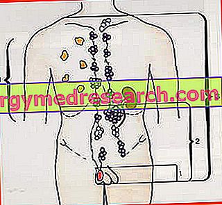
In a more than adequate number of patients, back pain, dull abdominal discomfort / pain and abnormal abdominal swelling coincide with the presence of lymph node metastases in the retroperitoneal area .
BILATERAL SEMINOMA
Seminoma generally affects a testicle only (unilateral). However, it can also be bilateral, ie affect both didymas.
Cases of bilateral seminomas are very rare and usually asynchronous. By asynchronous, we mean that tumor masses appear at different times and not simultaneously.
WHEN TO REFER TO THE DOCTOR
Not all palpable and painless testicular abnormalities, similar to those described, are seminomas (or a testicular tumor). Moreover, the latest statistical surveys report that less than 4% of testicular swelling has a neoplastic origin.
Nevertheless, doctors advise to undergo ad hoc examinations and checks in order to establish the exact nature of the problem. In fact, this is a completely precautionary measure.
COMPLICATIONS
If severe or untreated in time, a seminoma - like many other malignant testicular tumors and others - can spread to other parts of the body.
In fact, through the lymphatic and / or blood system, it can initially invade the neighboring lymph nodes and later the more distant lymph nodes, the lungs, the liver, etc.
This process is known as metastasis and the cancer cells that spread elsewhere are the so-called metastases .
Diagnosis
In general, the diagnostic procedure for the identification of a seminoma begins with the physical examination, continues with a scrotal ultrasound and blood analysis and ends with a biopsy.
If the tumor is in an advanced stage, it is likely that the doctor, in addition to the aforementioned diagnostic procedures, will also prescribe more or less invasive radiological checks.
EXAMINATION OBJECTIVE
During the physical examination, the doctor analyzes the testicles and in particular the one showing the swelling. Often, during the analysis, he also uses a small torch for the following reason: if the light passes through the bulge, it means that the latter contains liquid and is probably a non-malignant cyst; if instead the light does not filter, it means that the swelling is a solid mass and the solid masses generally have a neoplastic nature.
Once the testicles have been observed, the doctor checks the neighboring lymph node areas (in search of possible anomalies) and the distal ones (abdomen, neck, chest and armpits).
The physical examination also includes the investigation of clinical history, since, for diagnostic purposes, it is very important to know, for example, if the patient has a history of cryptorchidism, he comes from a family with a predisposition to seminoma, smokes etc.
SCROTAL ECOGRAPHY
Scrotal ultrasound is a non-invasive diagnostic procedure that allows, through an ultrasound probe, to observe the inside of the scrotum.
It provides a lot of useful information: it clarifies the position and size of the testicular anomaly, shows whether the swelling contains liquid or solid material, etc.
BLOOD ANALYSIS
Blood tests are used to track the so-called tumor markers in the bloodstream.
Tumor markers are substances that a tumor, when it appears and grows, can disperse in the circulating blood. In other words, they are a kind of distinctive elements.
Their identification is very significant for diagnostic purposes, however it should be noted that not all tumors produce them. Therefore, the absence of tumor markers does not always mean the absence of neoplasia.
Tumor markers present in case of seminoma:
- HCG (chorionic gonadotropin)
- LDH (lactate dehydrogenase)
- PLAP (placental alkaline phosphatase)
BIOPSY
A biopsy consists of the collection and histological analysis, in the laboratory, of a sample of cells from the suspected tumor mass.
Among those listed, it is the most reliable diagnostic test to establish the exact origin of the swelling, as well as, if it is a seminoma, its most important characteristics (gravity, stage of progress, etc.).
RADIOLOGICAL TESTS
Executable radiological examinations include chest radiography, nuclear magnetic resonance (NMR) and CT (computerized axial tomography).
The doctors resort to it when the diagnosis of seminoma is certain, in order to understand if the tumor has dispersed some metastases in the rest of the body.
GRAVITY OF A SEMINOMA: THE TUMOR STAGES
Only thanks to an accurate diagnosis, the doctor is able to establish the severity of a seminoma .
The parameters of evaluation of gravity - which is defined in stages - are the size of the tumor mass and the diffusion capacity of the tumor cells.
Based on these two parameters, a seminoma can be:
- Stage 1, if the tumor is limited to the affected testicle.
- Stage 2, if the tumor affects the affected testicle and the neighboring lymph nodes of the abdomen and pelvic area (retroperitoneal lymph nodes).

Treatment
The first and most important treatment to be applied in case of seminoma - as well as in the presence of any other testicular cancer - is the surgical removal of the entire testicle presenting the tumor mass. This surgery is known as inguinal orchiectomy .
If the stage of seminoma is superior to the first, removal of the diseased testicle is not sufficient, but requires integration with: surgical removal of the retroperitoneal lymph nodes and various cycles of chemotherapy and / or radiotherapy . The goal of these supplementary treatments is the definitive elimination of neoplastic cells from the body.
If the seminoma is bilateral, the inguinal orchiectomy is bilateral.
Note: if they consider it appropriate, doctors can resort to chemotherapy and radiotherapy even in the presence of stage 1 seminomas. In such circumstances, the purpose of these treatments is purely precautionary (adjuvant therapy).
INGUINAL ORCHIECTOMY
Performed under general anesthesia, the inguinal orchiectomy requires the doctor to make an incision in the groin, through which he can extract the entire ill testicle.
The removal of a testicle only (therefore when the seminoma is unilateral) does not affect the patient's libido or even fertility (ie the ability to have children). In fact, the remaining testicle makes up for the lack of the removed one, producing more testosterone and more sperms than normal. In other words, a process of hormonal and sperm compensation that protects fertility is triggered in a completely natural way.
From the point of view of results, these are better if the diagnosis is early and if the removal of the diseased testicle takes place when the neoplasm is at stage 1.
If the inguinal orchiectomy is bilateral
In the case of bilateral inguinal orchiectomy, the production of testosterone (which affects libido) and that of sperm (which affects fertility) are lost. If, due to the lack of testosterone, there is a solution - consisting in the administration of exogenous testosterone - there is no remedy for the absence of spermatozoa and the patient becomes sterile.
SURGICAL REMOVAL OF LYMPHONODES
It is also necessary to surgically remove the retroperitoneal lymph nodes, when the seminoma is at least at stage 2.
Although rarely, the operation of removing the lymph nodes can lead to a curable disorder known as retrograde ejaculation . This complication, which consists in introducing the ejaculate into the bladder, is due to damage to some nerve endings near the intervention area.
CHEMOTHERAPY AND RADIOTHERAPY
Chemotherapy consists of the administration of drugs (called chemotherapeutic drugs) capable of destroying all the rapidly growing cells, including cancer ones. In case of seminoma, the most prescribed chemotherapeutic drugs are bleomycin, etoposide and cisplatin.
Radiation therapy, on the other hand, involves exposing the patient to a certain amount of high-energy ionizing radiation, with the aim of destroying the neoplastic cells.
The number of chemotherapy and radiotherapy cycles depends on the stage of seminoma. This means that the more advanced a neoplasm is, the more therapeutic cycles are generally required.
| Main side effects of chemotherapy and radiotherapy. | |
| Chemotherapy | Radiotherapy |
| Nausea He retched Hair loss Sense of fatigue Vulnerability to infections | Nausea Sense of fatigue Diarrhea Skin redness Predisposition to other tumors |
Prognosis
If diagnosed and treated in time, seminoma - like many other testicular tumors - is one of the most likely malignant tumors with complete recovery .
In fact, according to the latest statistical surveys, the surgical treatment of seminomas at stage 1 allows the achievement of complete recovery in about 95% of cases. In other words, after the inguinal orchiectomy, they completely heal more than 9 out of 10 patients.
Obviously, this situation changes if the seminoma is more advanced (ie stage 2 or stage 3). In these situations, complete remission from the disease affects (at most) 70-75% of patients.
POST-THERAPEUTIC CHECKS
Despite the complete removal, the seminoma - like any malignant tumor - can reappear after some time ( recurrence ).
To promptly identify a recurrence of this testicular tumor, doctors plan a series of periodic checks, which the patient must follow scrupulously.
Generally, if the seminoma has not produced a recurrence during the first three years, it is rare for it to fail in the future.