Generality
Urethral stricture consists of the narrowing of the urethra, which is the channel through which urine accumulated in the bladder is expelled.

The main symptom consists in the difficulty in urinating, which, in the long run, can give rise to various complications, even serious ones.
The treatment of urethral stricture is based exclusively on surgery. In fact, there are no less invasive treatments able to reduce stenosis. However, it is comforting that the techniques used today provide satisfactory results.
What is the urethra?
To learn more: Urethra - Anatomy and Functions
The urethra is a tubular channel, which allows urine to flow outwards; originates from the bladder and ends with a small opening, called the urinary meatus.

Figure: urinary and male genital apparatus. Urethral stricture can affect both men and women, but is much more common in males.
The urethra is much longer in humans than in women; the male one, in fact, measures 18-20 cm and crosses the penis up to the tip of the glans; the female one, on the other hand, is much shorter and ends at the vulva (vestibule vulva), anterior to the vaginal opening and posterior to the clitoris.
In humans, the urethra also has another function: it is the way through which sperm passes during ejaculation.
What is urethral stricture
Urethral stricture consists in narrowing the urethra at any point in its path. The consequences of this shrinkage concern the flow of urine, whose passage is hindered; the affected person may therefore experience more or less severe urination difficulties.
The formation of a mass of scar tissue at the restricted area determines the urethra stricture. The greater this mass, the greater the occlusion of the urethral canal.
Epidemiology
The precise incidence of urethral stricture is not known. However, it has been observed that this disorder is much more widespread among males in their fifties. In fact, women and children represent only a small percentage of cases.
Furthermore, another important statistical fact concerns the causes. Today, urethral strictures from the Neisser gonococcus bacterium are decreasing, as a wide range of antibiotics has reduced the chances of contracting this infection and developing associated complications.
Causes
What determines the formation of the scar mass occluding the urethra? The causes of urethral strictures are different and concern:
- Traumas or injuries that damage the urethra
- Infections
- Congenital defects
- tumors
TRAUMAS AND ACCIDENTS
Accidental traumas or shocks to the urethra can damage this channel. When this occurs, the urethral lesions heal, creating around them a mass of scar tissue. In the most unfortunate cases, or if the trauma is significant, it may happen that the scar mass arrives to occlude the urethra.
The classic traumatological situations potentially responsible for the consequences described above are represented by falls (from cycles, motorcycles or horses) and from some surgical operations on the urinary tract (bladder and prostate) and genitals (hypospadias).
INFECTIONS
Scar tissue can also be formed following a pathogenic infection . This is the case of two infectious diseases, transmitted by sexual means, such as gonorrhea and chlamydia ; or infections caused by prolonged use of a urinary catheter; or, again, due to an inflammatory state of the prostate or the tissues surrounding the urethra.
It should be noted that an infection of the urethral canal does not always translate into a stricture. However, in its presence, the chances of this happening increase, especially when the pathology is neglected.
CONGENITAL DEFECTS
Despite being very rare, some children may be born with a congenital urethral canal defect.
TUMORS
Urethral tumors can narrow the urethral canal. However, this circumstance is also very rare.
Symptoms and complications
To learn more: Urethral Stenosis Symptoms
The symptoms of urethral stricture depend on the degree of stenosis itself. The less severe cases are asymptomatic, that is, they do not show obvious symptoms; vice versa, the most serious cases are characterized by increasingly severe disturbances. For example, the classic symptom of urethral stenosis, that is difficult urination, from a slight discomfort in mild cases, becomes a very serious problem in the most worrying cases.
The patient with urethral stricture complains:
- Reduced urine flow, the so-called reduced mite
- Painful urination
- Mitto to "spray"
- Drip shortly after urinating (post-voiding)
- Incontinence
- Incomplete emptying of the bladder
- Need to urinate often
- Urinary infections
- Blood in urine and seminal fluid
- Low jet ejaculation
DIFFICULTY ORINATION: CHARACTERISTICS
The main symptoms of urethral stricture concern the flow of urine. The patient experiences difficulties, and sometimes pain, already when urinating begins. The jet (mitto) is then reduced and the quantities of urine excreted are lower than normal. This explains why you feel the sensation of a missed bladder emptying and why you lose a few drops of urine, immediately after urinating.
Moreover, it may occur that the jet assumes the appearance of a spray ("mitto a spray") or that it is double.
COMPLICATIONS
A first complication, which characterizes urethral stricture, concerns the failure to empty the bladder after each urination. The repetition over time of this circumstance can turn into serious infections of the bladder, prostate and kidneys. Not by chance, the stagnation of urine inside the bladder is considered one of the main causes of infection of the urinary tract.
The other noteworthy complication concerns the mitto (urine jet) and the dimensions of the occlusion. In fact, if the conditions of a stenosis worsen, the patient could suffer from complete urinary retention, that is to say of total inability to urinate.
WHEN AND WHO TO CALL?
When the symptoms described are accompanied by pain and the general situation affects the standard of living, it is advisable to consult a specialist: the urologist .
Diagnosis
The diagnosis of urethral stricture involves numerous tests. Part of them serves to assess the severity of the disorder; another part helps the doctor understand the root cause. All this is essential to establish which therapeutic path is most appropriate.
It begins with a physical examination of urological evaluation and an investigation of the patient's medical history (medical history). We then proceed with a culture test of urine (urine culture ), a urethral swab and instrumental examinations, such as:
- Ultrasound of the urethra
- Retrograde urethrography
- Cistouretrography (or anterograde urethrography)
- Cystoscopy (or cysto-uretroscopy)
UROLOGICAL ASSESSMENT AND CLINICAL HISTORY.
During the urological examination, it is the duty of the physician to ask the patient about the symptoms experienced and his medical history, trying to go back to the time of appearance of the disorder. In fact, since a urethral stricture can be a direct consequence of a surgical operation or a fall from the bicycle, the fact of being aware of it represents, for the doctor, the first step towards a correct diagnosis.
Furthermore, it is equally important for the urologist to rely on a test that is easy to perform and not at all invasive: uroflowmetry . Through the latter, the amount of urine emitted per unit of time is measured, in other words the extent of urination. In patients with urethral strictures, the value, relative to the range, is lower than normal.
Uroflowmetry is a practical exam, but its limits should not be overlooked: low values also characterize other urinary tract diseases.
URINE CULTURAL EXAMINATION AND URETHRAL BUFFER
Urine culture and urethral swab are two tests aimed at finding particular pathogenic microorganisms, respectively, in the urine and in the low urinary tract. They are useful to the doctor if he suspects the presence of a bacterial infection in progress. The finding of certain bacterial strains (and of the relative infection) has important diagnostic meanings, since it allows:
- Go back to the causes of stenosis, such as, for example, in those cases due to gonorrhea and chlamydia
- Establish urine stagnation in the bladder
- Establish the most appropriate antibiotic therapy, based on the bacteria involved.
IN-DEPTH INSTRUMENTAL EXAMINATION
- Ultrasound of the urethra . This is a non-invasive radiological examination, for which no special preparation is needed. The use of a probe is foreseen, which the doctor runs along the affected area. It is very useful if the patient is a man, since the probe provides clear images relating to the degree of stenosis. Conversely, when the patient is a woman, the exam has little use and application.
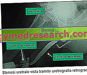
- Retrograde urethrography . It is also a radiological examination, which allows to analyze the integrity of the final tract of the urethral canal. The exam involves the injection, inside the urethra, of an iodinated contrast medium. This is done with a catheter. Once injected, the contrast medium flows along the urethral duct, adapting to the cavity it passes through. Therefore, if there are any restrictions, it penetrates inside and passes through them. The path taken by the contrast medium is revealed by a series of X-ray images.
- Cistouretrography or anterograde urethrography . It is a very similar examination, for execution, to the previous one. The only difference concerns the point of the urethra in which the contrast medium is injected: in this case, it is close to the bladder.
- Cystoscopy . It is an endoscopic examination of the urethral canal. An instrument, called a cystoscope, equipped with a camera is used. The cystoscope is inserted inside the urethra and, through a monitor connected to the instrument, the urethral lumen is observed. If there are injuries, anomalies and shrinkage, these are highlighted. Furthermore, it is also possible to take a small sample of tissue (biopsy).

Therapy
The therapy of urethral stenosis is based, mainly, on the surgical intervention and on the administration of antibiotics . If the latter are used to cope with bacterial infections of the urinary tract, surgery is the only countermeasure that can repair the damage of the urethral canal.
There are several possibilities for intervention:
- Urethral dilatation, by catheter
- The uretrotomia
- Surgical correction of the urethra
- The placement of a urethral stent (urethral stenting )
The choice of one procedure, rather than another, depends on several factors, such as: age, sex, general state of health of the patient, severity of stenosis and experience of the surgeon. The following table, instead, shows the clinical conditions, which oblige the operation.
When is it necessary to operate?
- Difficult to urinate
- Urinary retention
- Serious kidney and bladder problems
- Recurrent urinary tract infections
- Stagnation of urine in the bladder
- Unbearable pain
URETRAL EXPANSION
Urethral dilation is performed under local or general anesthesia, by inserting increasingly larger catheters into the urethra. Progressively increasing the diameter of the catheters serves to widen the narrowing gradually and not traumatically. The patient undergoes this operation several times. Repeating the intervention is fundamental for its success. In some cases, lubricating gels are used to facilitate the insertion of the catheters.
The uretrotomia
The urethrotomy uses an endoscope, equipped with a camera, which serves to recognize the exact point of the stenosis. Once the area has been identified, the urologist surgeon takes a tiny blade and performs the cutting and reopening of the occlusion. While waiting for this incision to heal, a Foley catheter (with an inflatable end) is introduced and left in place, only for a few days, in order to keep the urethral canal open.
We have seen that the success of the urethrotomy depends on the size of the stenosis. In fact, the smaller the size of the stenosis, the greater the chances of success; and viceversa.
SURGICAL CORRECTION OF THE URETHRA
The techniques of surgical correction of the urethra are different and depend on the size of the stenosis.
For small strictures, the surgeon first cuts and eliminates the area of scar tissue; after that, reconnect the two separate strips of urethra.
In the case of severe stenosis, instead, after removing the scar mass, a buccal tissue transplant is performed to reconstruct the missing urethra section.
Surgical correction has a good success rate. However it is an invasive procedure, to be performed under general anesthesia.
URETHRAL STENTING
Urethral stenting is another endoscopic procedure, such as uretrotomy. At the point where the urethra is restricted, a small tube, called a stent, is introduced, which serves to keep the channel open.
If the health conditions of a patient do not fit into any of the three previous operations, the stenting procedure is a valid alternative. This is the case, for example, of very old patients.
Prognosis
The prognosis of urethral stricture depends on several factors.
If left untreated, there is no possibility of healing. In fact, antibiotics are used to treat any bacterial infections, but all other symptoms remain.
Surgery therefore becomes a necessity.
The success of the operation depends on several factors. The main ones are:
- Age
- Severity of stenosis
- Surgical procedure best suited to the patient's health conditions
The outcome of the intervention is usually favorable. However, in more severe cases and in very elderly patients, relapses may occur that will require a second intervention to resolve urethral stricture.