The aorta is the main artery of the human body, both in size and in terms of elasticity: in the adult, it is approximately 30-40 cm long and has an average diameter of 2.5-3.5 cm.
The aorta originates from the heart, in particular from the left ventricle, which pushes inside it the oxygen-rich blood coming from the left atrium (where the pulmonary veins come out). The task of the aorta is therefore to distribute the oxygen-rich blood to the lower caliber arterial vessels; these, in turn, branch out repeatedly to vascularize the tissues of the entire organism. The aorta, however, is not a simple blood transport duct, but a real organ: thanks to the strong elasticity of its walls it is able to expand during systole and to relax during diastole, so as to ensure a constant blood flow in secondary arteries. The aortic endothelium also secretes numerous vasoactive peptides able to modulate the activity, not only of the various structures of the vessel wall, but also of the blood cells and of the coagulation system proteins that come into contact with it.
If we compare the heart to the roots of a tree, the aorta represents the trunk with its branches. All the arteries of the general circulation derive from the aorta.
Watch the video
X Watch the video on youtubeThe aorta is divided into two large segments:
|
Ascending aorta

About five centimeters long, the ascending aorta can be divided into two sections:
- aortic root : consists of:
- aortic or semi-lunar valve: formed by three cusps (tissue flaps), two posterior and one anterior, it opens during the left ventricular systole allowing the release of blood pushed into the aorta by contraction of the ventricle
- aortic sinuses of Valsalva: just above the origin of the aorta, there are three swellings, located behind the valvular cusps, which welcome the excursions of the valve flaps. Taken together, these dilations form a bulge called the bulb
- Anterior and posterior coronary hosts, from which two collateral branches originate respectively - the coronary of right and left - that carry the oxygen-rich blood to the myocardium
- tubular tract : extends to the aortic arch. At the junction with the aortic arch it is possible to recognize a more or less wide dilation on the right side, defined as a large aortic sinus, whose diameter is accentuated with age and can become the site of aneurysms
Aortic Arch
The aortic arch follows the ascending aorta. Starts in front of the trachea on the left and later on it also relates to the esophagus. It starts at the height of the upper margin of the second right sternocostal articulation; from here

From the aortic arch originate, from right to left:
- the brachycephalic arterial trunk (or anonymous artery) → dividing into the right common carotid artery and into the right subclavian artery, carries blood to the right arm, neck and head
- the left common carotid artery → carries blood to the neck and head
- the left subclavian artery → carries blood to the left arm
Sometimes, at the point where the aortic arch continues into the thoracic segment (corresponding to the sternal extremity of the second left rib cartilage) an annular narrowing can sometimes be noticed, to which the name of aortic isthmus has been given. This narrowing is immediately followed by a dilation, the so-called aortic spindle.
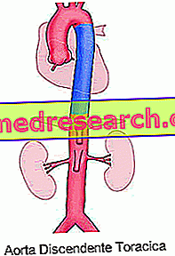

The descending aorta follows the aortic arch. It descends into the thorax through the posterior mediastinum, in front of and laterally to the vertebral column: it starts from the inferior margin of the IV thoracic vertebra and ends in front of the inferior margin of the XII thoracic vertebra, in correspondence of the diaphragmatic orifice.
From the thoracic aorta originate parietal branches that supply the thoracic wall and the diaphragm, and visceral branches that vascularize the organs contained in the thorax.
- Parietal branches: posterior intercostal arteries and superior phrenic arteries
- Visceral branches: bronchial arteries (supply the tissues in the lung), pericardial arteries (supply the pericardium), mediastinal arteries (the mediastinum) and esophageal arteries (vascularize the esophagus)
Abdominal Aorta
The abdominal aorta follows the thoracic aorta, begins in the diaphragm and runs parallel to and to the left of the inferior vena cava. It ends at the level of the body of the IV lumbar vertebra, where it bifurcates originating the two common right and left iliac arteries.

- Celiac tripod → vascularize the liver, stomach, esophagus, gallbladder, duodenum, pancreas and spleen
- Mesenteric arteries (upper and lower) → all together they vascular the small intestine, the large intestine and the pancreas; the superior mesenteric irrigates the pancreas, small intestine and the initial tracts of the large intestine, while the inferior mesenteric vascularizes the terminal portion of the colon and rectum
- Renal arteries → vascularize the kidneys
Furthermore, the abdominal aorta gives rise to the lower phrenic arteries (diaphragm and lower portion of the esophagus), to the adrenal arteries (the adrenal glands), to the renal arteries (the kidneys), to the genital arteries (testicular arteries in man and ovarian arteries). in women) and the lumbar arteries (vascularize the spinal cord and the abdominal wall).
The abdominal aorta continues inferiorly in the common right and left iliac artery - which are divided into internal and external iliac arteries vascularizing the pelvis and lower limbs - and ends with the middle sacral artery placed on the anterior aspect of the sacrum.
Summary table
Outline of Histology
Like all blood vessels, the aortic wall also consists of three overlapping tunas, which from the inside outwards are called:
- intimate habit: formed by an endothelium resting on a thin connective layer called a basal lamina
- medium cassock: formed mainly by an elastic connective component
- adventitious cassock: made up of connective tissue, it collects the vasa vasorum, that is the nourishing vessels for the arterial wall itself
Aortic disorders
- AORTIC ANEURISM: excessive and permanent dilation of the aortic lumen: it mainly affects smokers, diabetics, people with high blood pressure (hypertensive) and those with high values of cholesterol in the blood (dyslipidemic) and atherosclerosis; also some systemic diseases (Marfan syndrome) and some infections (syphilis) favor its onset
- AORTIC DISSECTION: the blood penetrates the middle layer of the aortic wall dividing it longitudinally and forming a false lumen; appears more easily at an underlying aortic aneurysm. Among the causes that favor the rupture of the vessels at the level of the average aorta, we recall: syndromes such as those of Marfan and Ehlers-Danlos, Noonan, Turner, congenital cardiovascular anomalies, inflammation, pregnancy, trauma, atherosclerotic ulceration, cocaine abuse, and iatrogenic causes for surgery or catheterization
- INTRAMURAL HEMATOMA: similar to aortic dissection, it is characterized by the absence of flow in the false lumen of the aorta.