Generality
The lobes of the brain are the 4 large sections that divide the cerebral cortex of each hemisphere of the brain and which are also known as frontal lobe, parietal lobe, temporal lobe and occipital lobe.

Protected by the homonymous bones of the neurocranium and characterized by convolutions and grooves, the lobes of the brain each have a specific set of functions; for example, the occipital lobe presides over the interpretation of visual stimuli, the parietal lobe to the processing of sensitive information coming from the skin, to the sense of position etc., the frontal lobe to the ability to produce spoken and written language, to the control of voluntary movements etc., and, finally, the temporal lobe, the ability to understand written and spoken language, to the perception and interpretation of sounds, etc.
Brief anatomical review of the brain
The brain is, together with the spinal cord, one of the two fundamental components of the central nervous system .
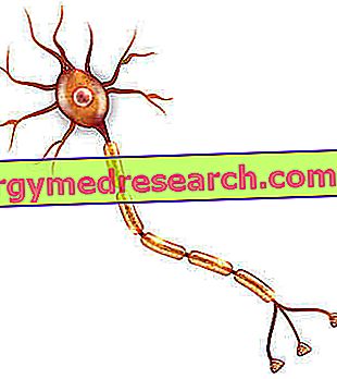
Heavy about 1.4 kilograms and containing 100 trillion neurons (in the adult human being), the encephalon is a very complex structure, which can be divided into 4 large regions, which are: the brain proper (or telencephalus or , simply, brain ), the cerebellum, the diencephalon and the brainstem .
BRAIN PROPERLY SAID
The brain is the largest and most important region of the brain.
Its general anatomical shape requires the presence of:

- Two large specular hemispheres (the right cerebral hemisphere and the left cerebral hemisphere ), separated by a groove (the so-called interhemispheric groove ), and
- A corpus callosum, located at the base of the two aforementioned cerebral hemispheres.
On the surface, the brain has the so-called gray substance, which goes to make up a laminar layer called the cerebral cortex ; in the deeper layers (therefore below the surface), instead, it presents the so-called white substance .
What are Brain Lobes?
The lobes of the brain, or cerebral lobes, are the 4 large sections, in which the cerebral cortex of each hemisphere of the human brain is ideally divided and which the anatomists have indicated with the names of frontal lobe, parietal lobe, temporal lobe and occipital lobe .
The purpose of this article is to describe the individual lobes of the brain, both anatomically and functionally.
Anatomy
The lobes of the brain reside inside the neurocranium (or cranial box ), that is the set of bones of the skull designed to protect the entire brain.
The lobes of the brain present a common architecture; each of them, in fact, possesses a series of ridges - whose specific name is convolutions - separated by more or less deep grooves - whose most appropriate name is furrows .
Frontal lobe
The frontal lobe is the lobe of the brain representing, in each cerebral hemisphere, the anterior portion of the cerebral cortex; the frontal lobe is therefore the cortical area of the brain that is located anteriorly to the remaining 3 cortical areas.
Mainly protected by the frontal bone (cranial bone that forms the forehead) and only for a small part by the parietal bone (cranial bone that constitutes the upper region of the cranial vault), the frontal lobe is, by extension, the largest of the lobes of the brain ; in fact, 41% of the entire cerebral cortex belongs to it.
Included within the so-called anterior cranial fossa, the frontal lobe is bordered by:
- The parietal lobe, posteriorly;
- The temporal lobe, posterolateral;
- The ipsilateral orbital cavity and the so-called anterior cranial fossa floor, below;
- Frontal bone, anteriorly;
- The frontal bone and part of the parietal bone, above.
To delimit its extension with respect to the neighboring lobes of the brain (parietal lobe and temporal lobe) are two deep grooves of the cerebral cortex: the so-called central sulcus (or Rolando sulcus ), as regards the boundary with the parietal lobe, and the so-called lateral fissure of Silvio (or fissure silviana ), as regards the border with the temporal lobe.

On the frontal lobe, several important functional areas of the brain are located; among these are: the primary motor cortex (on the precentral gyrus), the premotor cortex (on the precentral, upper frontal and middle frontal convolutions), the additional motor area (on the same convolutions of the premotor cortex), the Broca area ( on the inferior frontal gyrus) and the prefrontal cortex (on the remaining convolutions). On the frontal lobe, moreover, there is a very rich quantity of dopamine-sensitive neurons (NB: between the lobes of the brain, the frontal lobe is the one with the greatest quantity of these neurons).
Parietal lobe
The parietal lobe is the lobe of the brain constituting, in every hemisphere, the portion of the cerebral cortex between the frontal lobe, anteriorly, the occipital lobe, posteriorly, and the temporal lobe, inferiorly.
Protected by the parietal bone, the parietal lobe represents 19% of the entire cerebral cortex, which places it in third place in the special classification relative to the extension of the lobes of the brain.
The parietal lobe has well-defined boundaries: to separate it from the frontal lobe, there is the already mentioned groove of Rolando; to divide it from the temporal lobe, there is the already mentioned lateral fissure of Silvio; finally, to distinguish it from the occipital lobe, there is the groove known as the parieto-occipital sulcus .
Two important functional areas of the brain reside on the parietal lobe, which are:
- The primary somatosensory cortex (or primary somesthetic area ). To be precise, this functional area of the brain locates in the post-central convolution of the parietal lobe, ie the convolution between the Rolando sulcus and the post-central sulcus;
- The posterior cortical cortex . More in detail, this functional area of the brain is located in the convolutions of the superior parietal and inferior parietal lobules, convolutions that extend from the post-central sulcus, in the direction of the occipital lobe.
Temporal lobe
The temporal lobe is the lobe of the brain representing, in each cerebral hemisphere, the latero-inferior portion of the cerebral cortex.
Defended by the temporal bone (bone that includes the temple, the ear and the region immediately behind the ear), the temporal lobe covers an area of the cerebral cortex equal to 22% of the total, resulting in the second largest cerebral lobe after the frontal lobe.
Included within the so-called middle cranial fossa, the temporal lobe is bordered by:
- The parietal lobe, above;
- The frontal lobe, supra-anterior;
- The occipital lobe, posteriorly;
- The temporal bone, laterally;
- The floor of the middle cranial fossa, below.
The separation between the temporal lobe and the lobes of the parietal and frontal brain is clear, as it is marked by the presence of Silvio's often mentioned lateral fissure; the separation between the temporal lobe and the occipital lobe, on the other hand, is very blurred, as it lacks a deep and well defined anatomical groove (there is an imaginary line, called the parieto-temporal lateral line ).
On the temporal lobe take place the functional areas of the brain known as the Wernicke area, hippocampus and amygdala .
Occipital lobe
The occipital lobe is the lobe of the brain representing, in each cerebral hemisphere, the posterior portion of the cerebral cortex; in other words, therefore, it is the cortical area of the brain that develops posteriorly to the other 3 cortical areas.
Protected by the occipital bone (cranial bone of the anatomical region called occiput ), the occipital lobe covers an area of cerebral cortex equal to 18% of the total and this places it in the last place in the ranking relative to the largest lobes of the brain.

As part of the structures included in the so-called posterior cranial fossa, the occipital lobe borders:
- The parietal bone, anteriorly;
- The temporal bone, antero-laterally;
- The tentorium of the cerebellum, inferiorly;
- The occipital bone, posteriorly.
To delimit the area of extension of the occipital lobe are the aforementioned parieto-occipital sulcus, as far as the border with the parietal lobe is concerned, and the already mentioned lateral parieto-temporal line, as regards the border with the temporal lobe.
On the occipital lobe take place two important functional areas of the brain: the primary visual cortex (or calcarine cortex ) and the secondary visual cortex .
Curiosity: what is the tentorium of the cerebellum?
The tentorium of the cerebellum is the dura mater flap (one of the three meninges) which has the task of physically separating the cerebellum from the two occipital lobes; in a certain sense it is the anatomical structure that divides the cerebellum from the proper brain.
Brain lobes involved in the limbic system
The lobes of the frontal, temporal and parietal brain concur, with more intimate portions (and close to the underlying corpus callosum), to the formation of the so-called limbic system .
In neurology, the term "limbic system" refers to a complex of brain structures, which has a key role in emotional reactions, short-term memory processes, behavior and smell.
Did you know that ...
The functional areas amygdala and hippocampus, previously mentioned where the article dealt with the temporal lobe, are two components of the limbic system.
Function
The lobes of the brain each cover a specific set of functions . Despite this functional specificity, however, these brain areas are not at all disconnected structures; every lobe of the brain, in fact, is in communication with the others (and with other structures of the brain) and, indeed, its correct functioning depends on the correct functioning of the elements with which it is in contact (for example, the malfunctioning of the lobe frontal, induced by a lesion against him, can cause the malfunction of one or more of the other lobes of the brain).
The ability to see a color comes from the occipital lobe, while the ability to recognize it and identify it with a name depends on the temporal lobe.
The malfunctioning of one of these two lobes of the brain (it does not matter which one) always leads to an inability to establish the colors observed.
The lobes of the brain are not autonomous and independent organs, but fundamental components of that complex "machine" called encephalon.
In the next sub-chapters, the reader will be able to learn about the functions of the lobes of the brain and compare them with each other.
Frontal lobe: functions
The frontal lobe is important for:
- Control of voluntary movements . It is the prerogative of the primary motor cortex, of the premotor cortex and of the supplementary motor area;
- Long-term memory ;
- The production of spoken and written language . It depends on the presence of the Broca area;
- The ability to understand and react to the feelings of others ( empathy );
- The programming of behaviors and actions aimed at a certain result, to obtain gratification, to feel better, etc. ( reward system ). It is closely related to the dense presence of dopamine-sensitive neurons;
- The ability to plan, attention management (including selective attention) and impulse control . They are the prerogative of the prefrontal area;
- The ability to classify objects ;
- The personality .
Parietal lobe: functions
The parietal lobe has a key role in ensuring the sense of position and space and in the processing of sensitive information (such as pain, sense of heat or cold, touch, etc.) coming from the skin .
In addition, it contributes to memory capacity, computing skills and the ability to interpret language .
Temporal lobe: functions
The temporal lobe is the lobe of the brain that presides over:
- The perception of sounds, their recognition and their interpretation . To guarantee this is its close relationship with the components of the middle and inner ear;
- The interpretation of visual stimuli and the recognition, through the construction of a visual memory, of objects;
- Understanding of spoken and written language, and verbal naming and memory . These are functions that belong specifically to the Wernicke area;
- Long-term memory and control of seemingly unconscious functions, such as hunger, thirst, emotions, etc.
Occipital lobe: functions
The occipital lobe is the lobe of the brain responsible for the interpretation of visual stimuli ; to provide this capacity is the presence of the primary visual cortex and secondary visual cortex.
diseases
Due to head trauma, strokes, brain tumors and dementia, the lobes of the brain may be injured or altered in their normal anatomy; these injuries and alterations are the cause, as it is easy to understand, of their malfunctioning and of the loss, by the subject involved, of the functions under their control.
Symptoms of malfunctioning of the lobes of the brain
- The malfunctioning of the frontal lobe mainly induces: poor if not absent ability to control voluntary movements, expressive aphasia (inability to speak and write), abulia (loss of will), apathy, lack of empathy, personality changes, planning difficulties of strategies, judgments, behaviors or actions with a certain purpose and difficulty in controlling impulses.
- The malfunctioning of the parietal lobe is usually the cause of: loss of the sense of space, inability to recognize objects through touch (astereognosia uni-or bilateral), apraxia (inability to perform coordinated and directed gestures for a specific purpose), syndrome of Gerstmann (acalculia, digital agnosia, etc.), loss of sensory abilities, anosognosia (inability to recognize one's own deficits), loss of topographical memory, etc.

- The malfunctioning of the temporal lobe is primarily responsible for: receptive aphasia (inability to understand spoken and written language), acalculia (inability to calculate), agraphia (inability to formulate a written thought), verbal-auditory agnosia, dysphasia nominal, acquired dyslexia, quadrantonopsia (loss of a quarter of the visual field), alterations of non-verbal memory, prosopagnosia (inability to recognize people's faces) etc.
- The malfunctioning of the occipital lobe produces: hemianopsia (loss of half of the visual field), agnosia for the colors (lack of color recognition), acinetopsia (inability to see moving objects), visual hallucinations and Anton syndrome.
It is important to point out that, on the characteristics of the symptomatic picture deriving from the malfunctioning of the lobes of the brain, the extension of the lesion / triggering alteration and the involvement or not of the dominant cerebral hemisphere affect (eg: the lesions of the lobes of the brain) dominant hemisphere have more serious consequences of lesions of the brain lobes of the non-dominant hemisphere).