Generality
Dacryocystitis is an inflammation of the lacrimal sac.

The inflammatory process causes pain, redness, swelling of the tissue and excessive tearing. Furthermore, the digital pressure exerted on the lacrimal sac can cause purulent material to escape through the lacrimal dots. The most common complication is corneal ulceration.
The therapeutic management of dacryocystitis includes oral antibiotics, hot packs and dacryocystorhinostomy to repair the obstruction of the nasolacrimal duct.
Causes
Dacryocystitis is usually caused by an infection that begins in the tear ducts.
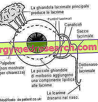
Dacryocystitis is determined by the narrowing or occlusion of the lacrimal tracts. If the tears are unable to drain, they accumulate in the lacrimal sac, thus becoming the cause of inflammation and excessive tearing of the eye (epiphora).
The pathological stasis of the tear fluid in the drainage system increases the risk of infection and makes the eyes more vulnerable to irritation.
Risk factors
Dacryocystitis is almost always associated with an obstruction of the nasolacrimal duct.
Factors that may increase the risk of developing the condition include:
- Stenosis for the growth of surrounding tissue;
- Injuries or traumas to the eye or adjacent tissues, infections, inflammations and neoplasms;
- Nasal disorders: deviation of the nasal septum, sinusitis, rhinitis, nasal polyps and hypertrophy of the nasal turbinates;
- Nasal or paranasal sinus surgery;
- Presence of dacriolites (white-yellowish calcareous formations) at various levels of the lacrimal drainage system, which determine mechanical obstruction.
Dacryocystitis can occur at any age, but tends to be more common in children. These, in fact, may also present a congenital obstruction of the nasolacrimal duct (defect referred to as dacrocistocele ).
Symptoms
To learn more: Symptoms Dacryocystitis
Dacryocystitis can occur suddenly (acute) or be long lasting (chronic). In chronic cases, tearing may be the only obvious symptom. In acute infection, the area around the lacrimal sac is painful, red and swollen. Furthermore, a slight pressure applied to the area can cause purulent material to escape through the opening of the lacrimal channels, in the inner corner of the eyelids (lacrimal dots).
Sometimes, a serious infection can cause the onset of fever and the collection of pus, which can also be discharged on the skin surface by forming a fistula. This closes, generally, after a few days of drainage.
Typical symptoms of acute dacryocystitis include:
- Inflammation: sudden onset of pain, redness and swelling in the area above the lacrimal sac, at the level of the medial chant of the lower eyelid, in the inner corner of the eye;
- Excessive tearing;
- Secretions of mucus or pus from the eye;
- Temperature.
If an infection of the nasolacrimal duct is not treated quickly or if it causes minor symptoms that accumulate over a long period, it may be more difficult to treat. Chronic dacryocystitis, in fact, shows less severe symptoms, but, over time, it can induce a further narrowing up to the occlusion of the tear ducts. Although epiphora and eye secretions may be present, pain is usually limited or absent, as is redness and edema.
In general, acute infections resolve rapidly with antibiotic therapy, while chronic infections, especially in adults, can be difficult to treat without surgery.
In newborns, tear canal obstruction is commonly self-resolving and exceeded at the age of 9-12 months.
Complications
The risks associated with untreated dacryocystitis mainly involve the risk of spreading infection on the surface (cellulite), deep (orbital, abscess or meningitis) or generalized (sepsis). These complications are rare and occur mainly in immunocompromised individuals.
Diagnosis of dacryocystitis
The doctor evaluates the presence of clinical signs that characterize dacryocystitis: swelling and redness in the inner corner of the eye, fever and excessive tearing. Pressure on the lacrimal sac can cause mucus or pus to escape. If purulent secretion is present, a sample can be taken and analyzed to determine which organism is causing the infection.
To confirm the diagnosis of dacriocystitis, the doctor can subject the patient to washing of the lacrimal passages, which allows to verify the presence of a complete or partial obstruction of the channels involved. A dye based on fluorescein is placed in the inner corner of the eye, so that it can flow into the tear film. If the tear drainage system is functioning properly, the dye should disappear from the surface of the eye after a few minutes.
The doctor can examine the puncture reflux by pressing on the tear channels and note any resistance. If structural abnormalities are suspected, dacryocystography and a CT scan of the orbit and paranasal sinuses can also be performed.
Treatment
If an obstruction of the tear duct is confirmed, in the absence of signs of infection, the doctor may recommend:
- Hot packs on the area (with a damp cloth);
- Gentle massages in the region of the lacrimal sac, to facilitate drainage.
In the event of a full-blown tear duct infection, the standard treatment is antibiotic therapy, which can be taken orally. These drugs can resolve acute infections rapidly and alleviate the symptoms of chronic dacryocystitis. However, if dacryocystitis does not respond to antibiotics and tends to recur, surgery may be necessary. Generally, the prognosis associated with surgery is good.
Different types of surgical treatments can be applied to dacryocystitis:
- Polling of the nasolacrimal duct, in which a thin wire is guided through the nasolacrimal duct to eliminate any blockage. This is the most common treatment for recurrent infections in newborns.
- In dacryocystorhinostomy, the reduced or obstructed nasolacrimal duct is expanded to prevent the infection from happening again. The procedure usually involves the creation of a discharge passage between the lacrimal sac and the nasal mucosa of the middle meatus, to prevent the accumulation of purulent material and allow the outflow of tears.