The renal glomerulus (from glomus, skein) is a dense spheroidal network of arterial capillaries, responsible for the filtration of blood.
The nephron
Each of the two kidneys of the organism contains about one and a half million nephrons. The nephron is considered the functional unit of the kidney, since on its own it is able to perform all the functions to which the kidney is in charge. Every single nephron can be divided into sections:
- Renal corpuscle: it is formed by the renal glomerulus and the Bowman's capsule; the latter is a hollow spherical structure with a blind bottom, which wraps around the glomerulus to collect the filtrate. On the whole, the renal glomerulus and Bowman's capsule constitute the renal corpuscle, also known as the Malpinghi or Malpighian corpuscle
- Tubular elements: the filtrate collected by the Bowman's capsule is channeled into a series of canaliculi, where it is deprived of useful substances for the organism (reabsorption) and enriched with those present in excess or considered dangerous (secretion). The continuous canalicular system is divided into three sections - proximal tubule, henle bend, distal tubule - each of which is specialized in the reabsorption and / or secretion of particular blood components
As explained above, the quantity of any substance present in the urine (excreted load) is the result of the following expression:
- Escreto load (E) = Filtered load (F) - Absorbed load (R) + Secrete load

For educational purposes, in the image above the nephron appears to be unfolded, when in reality it turns back and folds itself several times (image below).
The renal corpuscle
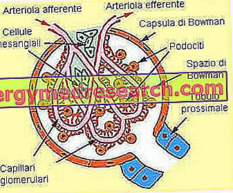
At the two ends of the renal glomerulus we find the two arterioles that put it in communication with the circulatory system. Upstream we find an arteriola, called afferent, which carries blood to be filtered; downstream we find an arteriola, called efferent, which conveys partially filtered blood into a network of capillaries distributed around the tubular elements.

As shown in the figure above:
- the afferent arteriola has a larger caliber than the efferent one.
- in juxtamidullar nephrons, the long peritubular capillaries that penetrate deep into the medullary area of the kidney are called vasa recta.
The blood drained from the peritubular capillaries is collected in venules and small veins that flow into the renal vein to convey the blood outside the kidney.
The renal glomerulus: what are its functions?
The renal glomerulus acts as a filter against the blood that passes through it.
Filtration is a passive, relatively non-specific process that marks the first stage of urine formation. As we will see better in the following chapter, the glomerular capillaries are called fenestrates, since they have relatively large pores through which many of the components of the blood can pass.

Overall, the volume of the renal ultrafiltrate is about 120-125 ml per minute, that is, equal to about 170/180 liters per day. Since the amount of urine is excreted more than 100 times lower, it is evident that the tubular system reabsorbs the vast majority of glomerular ultrafiltrate.
Along the tubular path, the ultrafiltrate undergoes a series of modifications that lead to a production of concentrated (definitive) urine equal to about 1 / 1.5 liters a day.
Filtration barriers
The blood is pushed by the hydrostatic pressure against the capillary walls of the glomeruli, favoring the passage of many of its components in Bowman's capsule, where they are collected by forming the ultrafiltrate (or pre-urine). To carry out this step, the blood components must pass through three different filtration barriers:
- the capillary endothelium: as anticipated, the glomerular capillaries are fenestrated capillaries, with large pores that allow most of the blood components to filter through the endothelium. The diameter of these pores allows the passage of many substances, resulting only too small for some plasma proteins and for blood cells (in the whole defined as corpuscolated elements), which remain in the blood. In particular, under normal conditions the fenestrated capillaries allow the filtration of molecules with a diameter less than 42 Å. Although the albumin molecule is smaller (36 Å), under normal conditions it cannot cross the capillary endothelium because it is blocked by negatively charged fixed proteins that repel it (albumin being also negatively charged).
- the basal lamina: the fenestrated endothelium of the blood capillaries rests on a thin basal lamina, called dense lamina, which separates the capillary endothelium of the bowman's capsule. The basal lamina consists of glycoproteins and a material similar to collagen (proteoglycans); both components are negatively charged, thus helping to repel most of the plasma proteins preventing their filtration
- the epithelium of Bowman's capsule: it contains specialized cells called podocytes (from podos, foot); each podocyte is characterized by cytoplasmic extensions, called pedicels, which protrude like tentacles from the cellular body wrapping the glomerular capillaries and resting directly on the dense lamina of the capillary wall. Thus, filtration cracks (slit pores) are formed, bounded by a membrane.
Similar to mesangial cells, the podocytes also have contractile fibers connected to the basement membrane by proteins called integrins. The contractility of these cell types is influenced by the endocrine action of certain hormones that regulate blood pressure and the balance of fluids in the body.

Thanks to these three barriers, the filtration of blood components results:
- free for molecules with radius <20 Å
- variable for molecules of radius 20-42 Å (70 - 150 Kd): the filterability between 20 Å and 42 Å depends on the charge. Since most of the plasma proteins have a negative charge, the filtration barrier strongly prevents or limits the filtration of proteins with a radius of 20-42 Å.
- absent for molecules radius> 42Å