Generality
The muscles of the thigh are the muscular elements that cross the anatomical section occupied by the femur, ie the bone constituting the thigh.
To simplify the study, anatomists divide the thigh muscles into two groups: the thigh muscles of the anterior compartment, the thigh muscles of the medial compartment and the thigh muscles of the posterior compartment.
The thigh muscles mainly allow hip flexion, leg extension, adduction movement of the lower limbs and hip extension.
Brief anatomical reference to the muscles
The muscles of the human body have two ends: an initial or proximal call and one called terminal or distal .
At each end, there is a tendon . A tendon is a formation of fibrous connective tissue, which connects a muscle to a bone element.
Therefore, the muscles find insertion on the skeleton by means of the tendons.
The anatomical texts and experts have a tendency to identify the initial extremity and the terminal extremity of a muscle with the tendon present on each of these extremities.
In anatomy, proximal and distal are two terms with the opposite meaning.
Proximal means "closer to the center of the body" or "closer to the point of origin". Referring to the femur, for example, it indicates the portion of this bone closest to the trunk.
Distal, on the other hand, means "farther from the center of the body" or "farther from the point of origin". Referred (always to the femur), for example, it indicates the portion of this bone furthest from the trunk (and closer to the knee joint).
Definition of thigh muscles
The muscles of the thigh are the muscles whose fibers take place, totally or only partially, in the anatomic-skeletal section formed by the femur ; the femur is the thigh bone.
The fact that the aforementioned muscles reside in the anatomic-skeletal section constituted by the femur does not necessarily imply their link with the bone in question; in other words, in the thigh there are muscles or parts of them, which do not interact in any way with the femur.
BRIEF DEFINITION OF THIGH
The thigh is the anatomical region of the human body lying between the pelvis, proximally, and the leg, distally.
On the border between pelvis and thigh, there is a very important articulation of the human body: the hip joint . On the border between thigh and leg, however, there is the knee joint, which is also very important and resulting from the interaction between the tibia (one of the two bones of the leg) and the femur.
BRIEF ANATOMICAL RECALL OF THE THIGH
To understand the placement of the thigh muscles, it is essential to bring to the attention of readers some anatomical features of the femur.
In the human being, the femur is the even bone that makes up the skeleton of the thigh . It belongs to the category of long bones and takes part in the formation of two important joints: the hip joint (femur-iliac bone) and the knee joint (femur-tibia).
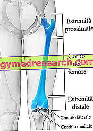
Like all long bones, the femur can be divided into three main portions: the proximal end (or proximal epiphysis ), the body (or diaphysis ) and the distal end (or distal epiphysis ).
- The proximal end of the femur is the bone portion located closest to the pelvis and involved in the constitution of the hip joint. At the proximal end, there are at least 6 regions of a certain anatomical relevance: the head, the neck, the great trochanter, the small trochanter, the anterior intertrochanteric line and the posterior trochanteric crest.
- The body is the central portion of the femur, included between the proximal end and the distal end. Similar to an hourglass, the body of the femur has a bony crest, called the sour line, which becomes the protagonist of a double bifurcation, an upper and a lower one. The upper bifurcation gives rise to the so-called pectineal line and to the so-called gluteal tuberosity. The lower bifurcation, instead, leads to the formation of the so-called lateral supracondylar line and of the so-called medial supracondylar line. The medial supracondylar line terminates its path with a protuberance, known as the adductor tubercle.

From the functional point of view, the femur is a fundamental bone for the equitable distribution of forces and body weight on the lower limb and for locomotion (the muscles that engage and the joints to which it takes part are essential for walking, running and jump).
In anatomy, medial and lateral are two terms of opposite meaning, which serve to indicate the distance of an anatomical element from the sagittal plane . The sagittal plane is the anteroposterior division of the human body, from which two equal and symmetrical halves are derived.
Mediale means "near" or "closer" to the sagittal plane, while lateral means "far or" farther "from the sagittal plane.
Anatomy
The anatomists divide the thigh muscles according to their location; it follows that they exist: the muscles of the thigh of the anterior compartment, the muscles of the thigh of the medial compartment and the muscles of the thigh of the posterior compartment .
MUSCLES OF THE THIGH OF THE FRONT COMPARTMENT
Located on the front of the thigh, the thigh muscles of the anterior compartment are a total of 4: the sartorius muscle, the pectineus muscle, the quadriceps femoris muscle and the ilio-psoas muscle.
- Sartorius muscle . The sartorius is the longest muscle in the human body and the most superficial of the anterior compartment. It is thin and crosses the entire thigh with an infero-medial orientation (ie downwards and towards the sagittal plane). It contributes to the formation of the so-called femoral triangle (or Scarpa's triangle ).
The sartorius muscle is an example of a thigh muscle that has no relation to the femur.
Proximal end: originates at the level of the anterior superior iliac spine . The anterior superior iliac spine is a characteristic prominence of the iliac bone .
Distal end: attaches to the upper medial surface of the tibia.
Innervation: up to the crural nerve (or femoral nerve ). Having both motor function and sensory function, the crural nerve is an important nerve of the peripheral nervous system (SNP) and represents the most voluminous branching of the so-called lumbar plexus. The lumbar plexus is an important reticular formation of various spinal nerves (which are also their nerves in the peripheral nervous system), which have the task of innervating part of the abdomen and the lower limbs in full.
Spraying: it is up to the femoral artery . The femoral artery is the most important arterial vessel in the lower limbs.
- Pectineus muscle . The pectineus is a flat and quadrangular muscle, located at the base of the femoral triangle. It is close to the muscles of the thigh of the medial compartment.
Proximal end: originates at the comb crestal level of the pubis (or pubic bone ).
Distal end: it is inserted at the level at the line of the femur, just below the small trochanter.
Innervation: it is up to the crural nerve and, in some individuals, to a branch of the obturator nerve.
Spraying: it is up to the obturator artery . The obturator artery is a branch of the internal iliac artery.
- Quadriceps femoris muscle . The femoral quadriceps is, in reality, a set of four different muscles: the vastus lateralis muscle, the intermediate broad muscle, the vastus medialis muscle and the rectus femoris muscle . The figure below is useful to understand what the real arrangement of the aforementioned muscular components is.
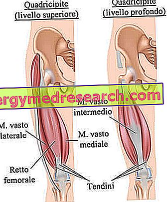
Proximal extremity: the vastus lateralis muscle originates, in part, at the level of the great trochanter and, in part, at the level of the sour line. The intermediate broad muscle originates from the anterior and lateral surfaces of the body (or diaphysis) of the femur. The vastus medialis muscle originates, in part, at the level of the anterior intertrochanteric line and, in part, at the level of the rough line. Finally, the rectus femoris muscle originates at the level of the ilium (one of the three bony portions that make up the iliac bone, together with ischium and pubis).
Distal extremity: the distal ends of all four muscles that make up the quadriceps femorate flow into a very large tendon, known as the patellar tendon . The patellar tendon crosses the knee patellar superiorly and is inserted at the level of the tibial tuberosity (or tuberosity of the tibia).
Innervation: up to the crural nerve.
Spraying: it is up to the femoral artery.
- Ilio-psoas muscle . The ilio-psoas is a muscle resulting from the union of two muscle elements: the large psoas muscle and the iliac muscle . The particularity of these two muscular elements constituting the ilio-psoas is the fact that, at their point of origin, they are two separate muscles and without any relation between them; while, in their terminal extremity, they form a whole.
Proximal end: the proximal end of the large psoas originates at the level of the lateral surface of the bodies of the vertebrae T12, L1, L2 and L3.
The proximal end of the iliac muscle, on the other hand, takes origin at the level of the so-called iliac fossa (which is a characteristic region of the ilium).
Distal end: it is inserted at the level of the small trochanter of the femur.
Innervation: the innervation of the large psoas is due to the branching of the spinal nerves L1, L2 and L3; the innervation of the iliac muscle, on the other hand, belongs to the crural nerve.
Spraying: it is up to the so-called medial circumflex artery of the femur and to the lumbar artery.

MUSCLES OF THE THIGH OF THE MEDIUM COMPARTMENT
Located in the inner portion of the thigh, the muscles of the thigh of the medial compartment are 5 in all: the gracilis muscle, the external obturator muscle, the short adductor muscle, the long adductor muscle and the large adductor muscle .
- Gracilis muscle . Among the thigh muscles of the medial compartment, gracilis is the most superficial and medial muscle. Thin and flattened, it crosses the hip and knee joints.
Proximal end: originates at the level of the so - called ischio-pubic branch . The ischio-pubic branch represents the point of union between the pubis and the ischium.
Distal end: it is inserted at the level of the medial surface of the tibia, precisely in the goose leg .
Innervation: it is up to the obturator nerve . The obturator nerve is a peripheral nerve, which derives from the lumbar plexus. Several very important nerve branches originate from the obturator nerve.
Spraying: it is up to the so-called medial circumflex artery of the femur.
- External shutter muscle . The external shutter is a flat and triangular muscle. Among the muscles of the thigh of the medial compartment, it is the smallest and the one located more on the surface.
Proximal end: originates at the level of the membrane that covers the so-called obturation hole.
Distal end: it is inserted at the level of the so-called trochanteric fossa of the femur . The trochanteric fossa of the femur is a small depression located near the large trochanter.
Innervation: it is up to the obturator nerve.
Spraying: it is up to the obturator artery.
- Short adductor muscle . The short adductor is a small muscle, which lies, for the most part, below the long adductor muscle.
Proximal end: it originates at the level of the anterior surface of two characteristic areas of the pubis, which are: the lower branch and the body.
Distal extremity: it is partially inserted at the level of the small trochanter and, partly, at the level of the rough line of the femur.
Innervation: it is up to the obturator nerve.
Spraying: up to the deep femoral artery.
- Long adductor muscle . The long adductor is a long, large and flat muscle. For a stretch of its path, it covers the short adductor muscle and the large adductor muscle.
The long adductor muscle contributes to the formation of the medial border of the so-called femoral triangle.
Proximal end: it originates in the body of the pubis.
Distal end: it is inserted at the level of the rough line of the femur.
Innervation: it is up to the obturator nerve.
Spraying: up to the deep femoral artery.
- Adductor large muscle . The large adductor is a triangular-shaped muscle, located deep beneath all the other muscles of the thigh of the medial compartment.
Anatomists often tend to recognize two components in the adductor muscle: a pubofemoral component and an ischiochondylar component .
The aforementioned components originate and end at different points.
Proximal end: the proximal end of the pubofemoral component originates, in part, at the level of the inferior branch of the pubis and, partly, at the level of the inferior branch of the ischium. The proximal end of the ischiochondylar component, on the other hand, originates from the ischial tuberosity.
Distal end: the distal end of the pubofemoral component is inserted at the level of the rough line of the femur. The distal end of the ischiochondylar component is inserted at the level of the medial condyle of the femur, precisely in the so-called adductor tubercle of the femur.
Innervation: it is partly due to the obturator nerve and partly to the tibial nerve.
Spraying: up to the deep femoral artery.
MUSCLES OF THE THIGH OF THE REAR COMPARTMENT
Located on the back of the thigh, the thigh muscles of the posterior compartment are altogether 3: the biceps femoris muscle, the semitendinosus muscle and the semimembranosus muscle .
The thigh muscles of the posterior compartment are also known by the English term hamstring .
- Hamstring muscle . The biceps femoris is a muscle that has, in the tract of origin, two heads (or heads), known as long head (or long head) and short head (or short head). Due to the presence of these two heads, the biceps femoris muscle is comparable to the biceps brachial muscle of the arm.
Proximal end: the long head originates at the ischial tuberosity of the ischium. The short head, on the other hand, originates at the level of the rough line of the femur.
Distal end: it is one and fits into the so-called fibula head.
Innervation: the innervation of the long head is due to the tibial component of the sciatic nerve, while the innervation of the short head belongs to the fibular component (ie the fibula) of the sciatic nerve.
The sciatic nerve is the largest and longest nerve in the human body. It begins, in fact, at the level of the back, along the whole lower limb, covering both motor functions and sensitive functions, and ends in the foot.
Spraying: up to the deep femoral artery
- Semitendinosus muscle . The semitendinosus is a superficial muscle, which covers, for the most part, the semimembranosus muscle.
Proximal extremity: it originates at the ischial tuberosity of the ischium.
Distal end: inserts at the medial surface of the tibia, precisely in the so-called goose leg.
Innervation: up to the tibial component of the sciatic nerve.
Spraying: it is up to the inferior gluteal artery.
- Semi-membranous muscle . The semimembranosus is a flattened muscle, located below the semitendinosus muscle. It is the most medial of the muscles constituting the hamstrings .
Proximal end: it originates at the ischial tuberosity of the ischium, but not exactly at the same point where the proximal end of the semitendinosus muscle originates.
Distal end: inserts at the medial tibial condyle.
Innervation: up to the tibial component of the sciatic nerve.
Spraying: it is up to the deep femoral artery and to the gluteal artery.

Function
The thigh muscles of the anterior compartment mainly allow the extension of the leg in the direction of the knee and flexion of the hip.
The thigh muscles of the medial compartment mainly allow the adduction movement of the lower limb. Lower limb adduction means the ability to bring the lower limb closer to the sagittal plane.
Finally, the thigh muscles of the posterior compartment mainly allow for hip extension and knee flexion.
For a complete picture of the functions performed by the thigh muscles, the table below is reported.
Function of the thigh muscles | |
Front compartment | Sartorium: allows flexion, abduction and lateral rotation of the hip. Comb: allows adduction and flexion of the hip joint. Femoral quadriceps: allows the extension of the leg in the direction of the knee and flexion of the hip. Ilio-psoas: supports flexion and lateral rotation of the hip. |
Medial compartment | Gracile: allows the adduction of the thigh and flexion of the leg. External shutter: allows lateral rotation of the thigh. Adductor short: allows the adduction of the leg. Adductor long: allows adduction and medial rotation of the thigh. Great adductor: allows the adduction of the thigh. |
Rear compartment | Hamstring: allows knee flexion and lateral extension and rotation of the leg, compared to hip and knee. Semitendinosus: allows flexion of the leg and extension of the thigh in the direction of the hip. Furthermore, it allows the medial rotation of the thigh with respect to the hip and the medial rotation of the leg with respect to the knee. Semimembranosus: allows the flexion of the leg and the extension of the thigh in relation to the hip. Furthermore, it allows the medial rotation of the thigh with respect to the hip and the medial rotation of the leg with respect to the knee. |
Associated pathologies
Like most of the muscles of the human body, the thigh muscles can also undergo contractures, strains, tears and inflammations / injuries in the tendons.
These injuries usually affect active people, such as those who practice sports.
ACCIDENTS TO HAMSTRING
The muscles that make up the so-called hamstring are particularly prone to injuries, especially among those who practice sports such as running, football, football or rugby.
The most serious problems to the thigh muscles of the posterior compartment are a source of intense pain and hematoma.