The evaluation of the conditions of the cardiovascular system constitutes the crucial moment of the visit to which every subject who practices sports, both competitive and not, is subjected. When irregularities are detected (eg puffs or electrocardiographic alterations), it is necessary to establish whether this finding should be considered physiological or pathological. If this last hypothesis occurs, the task of the sports doctor must be to be able to assess [using, in addition to the physical examination, also a series of instrumental examinations (electrocardiogram, phonocardiogram, telecuore, echocardiogram)] if the state pathological may give rise to deterioration, or if it may in some way expose the subject to sudden unforeseen events, such as death or syncope, dangerous both for the subject in question and for those who are having to witness such conditions.
It is also necessary that the evaluation takes place taking into account the particular type of sport that the subject intends to practice; in other words, the commitment of the cardiovascular system must be considered in that particular type of sport.
ELECTROCARDIOGRAM
Using the electrocardiograph it is possible to record, using appropriate electrodes, the electrical stimuli and transform them into a graphic signal: the electrocardiogram. The paper on which an electrocardiogram is recorded is millimetric: in a horizontal sense each square corresponds to 0.04 sec; each series of five small squares, delimited by a slightly more marked line, therefore lasts 0.2 sec. The duration of each electrical event is measured horizontally; on the other hand, the amplitude of the waves is measured vertically: 1 cm corresponds to 1 millivolt.
The currents that excite the heart are the result of a complex ionic movement (in particular of the ions, sodium, potassium, calcium, chlorine) that occurs between the intracellular and extracellular environment.
An electrocardiogram is formed by a series of waves and strokes that repeat cyclically; the sequence of the electrocardiographic elements that make up an electric cardiac cycle is the following: P wave - PR segment - QRS complex - ST segment - T wave - eventual U wave.
The P wave corresponds to the depolarization of the atria, or to the propagation of the electrical impulse from the sino-atrial node, where it is formed, to all the atrial musculature which consequently contracts; the electrical phenomenon precedes the mechanical phenomenon (ie the contraction). While in rest conditions the P wave has visible limits of duration and amplitude, in the subject under stress these limits can be far exceeded.
The PR tract is measured from the beginning of the P wave to the beginning of the QRS complex, ie the time taken by the electrical stimulus to activate the atria and pass through the atrioventricular node. In the normal subject its duration is between 0.12 and 0.20 sec, in the cross-country skiers it is greater.
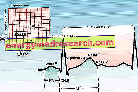
The QRS complex is the expression of the depolarization of the 2 ventricles; it also has limits in terms of duration and amplitude. As for the duration, it should not exceed 0.08 sec; as far as amplitude is concerned, the limits are much more inaccurate. However, the increased width of the QRS complex was found in the athlete.
Finally, the ST tract represents the repolarization of the ventricles.
The electrocardiogram can also be recorded when the subject produces an effort, pedaling on a cycle ergometer, or walking on a conveyor belt. These recordings are used to assess any changes to the electrocardiogram at rest (doubt of ischemia), or arrhythmias, or when one wishes to observe cardiac performance during muscular work.
phonocardiogram
The phonocardiogram transforms into a graphic signal the noises produced by the heart during its activity. Usually an electrocardiographic trace is also recorded simultaneously, so as to be able to correlate, with precision, mechanical events with electrical ones.
This examination is recorded by affixing a special probe to the chest, which is subsequently moved to the various auscultation foci. For each outbreak, multiple recordings are made, selecting different sound frequencies. The normal noises produced by the heart are the 1st and 2nd heart sounds. The 1st tone is produced by the closure of the atrioventricular valves; the 2nd tone is produced instead by the closing of the semi-lunar valves (aortic and pulmonary). Frequently, especially in young athletes, there is a physiological doubling of the 2nd tone, or the presence of a tone added at the beginning of diastole.
The intervals between the 1st and 2nd tone (systolic pause) and between the 2nd tone and the 1st successive tone (diastolic pause) are normally silent, but, in some cases they may present noises (murmurs) that will be called systolic or diastolic according to the pause they will occupy.
The phonocardiogram is used to assess a cardiac murmur more accurately; it will therefore be possible to establish precisely in which part of the cardiac cycle the breath is located, its intensity and frequency, and the particular morphology. All these elements are useful to distinguish the so-called innocent or functional murmurs, from those that derive from a heart disease. It is, however, an examination that is used much less frequently than in the past and that usually adds little to an accurate auscultation with the phonendoscope.
TELECUORE
It is the survey carried out using X-rays. The distance of the subject from the source of rays must be about 2 m in order to prevent the excessive divergence of the rays from causing distortions or enlargements of the structures whose images would be altered.
Due to the shape of the heart, it is usually not sufficient to make a projection in the anteroposterior sense, but it is necessary to make oblique and lateral projections (oblique anterior left and right, lateral-lateral). While in the antero-posterior projection the contrast between the transparency of the pulmonary fields and the cardiac shadow is sufficient, in the oblique and lateral projections it is no longer so it is necessary to ingest a radiopaque substance which, opacifying the esophagus, makes it evident on of it the imprint of any enlarged heart structures. In the normal subject, the heart can take on different radiological aspects, linked to the biotype, which explain the currently used terminology: horizontal heart (in the short one), oblique (in the normal type) and vertical (in the long line). Through special calculations, it is possible to obtain the measurement of the cardiac volume starting from the radiographic images. There is no doubt the interest of this datum, in particular in the evaluation of athletes: unfortunately the precision of the data obtained is not very high, due to some difficulties (such as the need to run the slab always in the same phase of the cardiac cycle, in so as to obtain comparable results) difficult to overcome. Moreover, in the same subject, the results obtained present considerable variability.
To obtain the cardiac volume, measurements are taken which are taken in anteroposterior projection (height and width of the cardiac shadow) and on the lateral projection (depth), obtained from the subject in horizontal decubitus position, since in this position there are fewer volumetric variations .
Finally, the Rorher formula is applied: cardiac surface x maximum depth x 0.63, which becomes 0.4 x length x width x maximum depth in cm.
It should be remembered that from normal values of 700-800 ml of volume, in athletes of endurance sports can reach about 1400 ml.
ECHOCARDIOGRAM
Physically, this type of investigation is based on a reflected ultrasonic beam that is picked up by a probe (the same one that emits the ultrasonic beam) and transformed into an electrical signal which, in turn, is converted into a graphic form, giving rise to images that correspond to the various structures of the heart in movement (the free walls of the ventricles, the septa, the valves, the cavities).
Echocardiography can be performed with a one-dimensional or two-dimensional technique. In the first case (one-dimensional technique), an isolated sector of the heart is explored; the spatial resolution is very good and it is possible to carry out a whole series of measurements concerning the dimensions of the ventricles, those of the atria, the amplitude of the valve movements and the quality of these movements. The two-dimensional technique gives us a complete view of the heart in motion, clarifying the spatial relationships between the various structures. The power of resolution is, however, less than in the one-dimensional technique.
In conclusion, it can be stated that the techniques described above should not be applied separately, but both are part of a complete echocardiographic examination.
Echocardiographic examination allows:
- accurately analyze the movements of all cardiac structures;
- perform rather precise measurements of the size of cardiac structures, evaluating the relationships existing between them;
- resolve any diagnostic doubts.
Echocardiography allows us to study the adaptation of the heart to different types of sports. In athletes dedicated to endurance sports, the main modifications concern the diameters of the cardiac cavities, which are also considerably increased, while the thickening of the walls is only moderate. These alterations, induced by training, are reversible within 2-3 months, if training is suspended. In athletes dedicated to power activities, an increase in the thickness of the ventricular walls occurs above all.
Edited by : Lorenzo Boscariol