Premise
In general, stomach cancer is an asymptomatic malignant neoplasm in the very early stages, therefore difficult to detect, even by a doctor with considerable experience.
As it progresses, the symptoms and signs gradually become more and more evident and, at this point, its diagnosis becomes easier (NB: in the table below is a summary of the typical symptomatic picture of stomach cancer).
Before proceeding further, readers are reminded that this article focuses on the diagnosis of a specific stomach tumor: gastric adenocarcinoma .
Also known as adenocarcinoma of the stomach, gastric adenocarcinoma is the best known and most common malignant neoplasm of the stomach (9 cases out of 10 of gastric cancer are gastric adenocarcinomas).
Typical symptomatological picture of gastric adenocarcinoma
- Discomfort in the epigastrium
- Dyspepsia
- Dysphagia
- Episodes of indigestion
- Stomach ache
- Nausea and vomit
- belching
- Weight loss
- Digested blood in faeces (melena or occult blood in stool)
- Hematemesis
- Anorexia, which sometimes becomes repugnance, for certain foods
- Recurrent fatigue
- Pain behind the sternum
- Presence of a swelling at the epigatric level
- Iron deficiency anemia
Diagnosis
The diagnosis of stomach cancer is often the result of a long process that begins with physical examination and medical history, proceeds with some laboratory tests on blood and faeces and, finally, ends with various instrumental tests and a biopsy.
Each step mentioned above is fundamental to the formulation of a correct and accurate diagnosis.
Physical examination and medical history
Physical examination and anamnesis are two diagnostic evaluations that provide useful information regarding the symptomatology (eg: they lead to the finding of discomfort in the epigastric area, dyspepsia, dysphagia, heartburn, food regurgitation, etc.).
Furthermore, they allow the doctor to understand the patient's general health and hypothesize the possible reasons for the symptomatic situation in progress (the anamnesis, for example, foresees an investigation relating to the risk factors related to a given symptomatic picture).
Although important, what emerges from the physical examination and the history does not allow us to formulate any definitive diagnosis; it is for this reason that more in-depth research is needed, such as laboratory tests and instrumental tests.
The investigations that characterize, generally, the physical examination and the anamnesis of a suspected case of stomach tumor.
- Measurement of blood pressure, heart rate and body temperature;
- Questions related to the characteristics of food digestion (in practice, how it happens and if it is problematic);
- Questions related to the presence of digestive disorders, such as diarrhea, constipation, vomiting, heartburn, epigastric pain, etc .;
- Questions intended to clarify whether there was an unexplained drop in body weight;
- Palpatory examination of the abdomen, in search of possible swellings in the epigastrium and / or liver;
- Observation of skin color;
- Questions related to a possible repugnance to certain foods, meat in particular.
Laboratory tests
Also useful but not sufficient for the formulation of a definitive diagnosis of stomach cancer, laboratory tests generally consist of:
- Blood tests,
- Faeces analysis e
- Quantification of tumor markers.
BLOOD TESTS
Blood tests show sideropenic anemia, a fairly important and common consequence in stomach cancer (50% of cases).
In addition, they provide:
- Information on albumin levels, a plasma protein whose concentration may decline in the presence of a sick stomach and unable to absorb proteins;
- Details of renal (ie kidney) and hepatic (ie liver) function.
ANALYSIS OF FECI
The analysis of the faeces allows to identify possible traces of digested blood in the faeces not visible to the naked eye (in substance, the so-called occult blood in the faeces ).
As a rule, the search for traces of digested blood in the stool is positive in almost 50% of clinical cases.
QUANTIFICATION OF TUMOR MARKERS
In medicine, those substances found in the blood which, in the presence of a neoplasm, are found in high concentrations take the name of tumor markers .
Tumor markers generally have a protein nature.
During the search for a stomach tumor, the tumor markers under observation are three: the CEA (or Carcino-Embryonic Antigen ), the alpha-fetoprotein and the CA 19-9 (or GICA, which stands for Cancer Antigen Gastro- Intestinal ).
It should be pointed out that, unfortunately, the finding of these tumor markers is almost always useless for the purposes of an early diagnosis: in fact, assuming that they vary in terms of concentration, CEA, alpha-fetoprotein and CA 19-9 increase in quantity only when the neoplasm gastric is in an advanced stage and has already metastasized.
- CEA or Carcino-Embryonic Antigen . Its finding at high levels occurs in 40-50% of patients with advanced and metastasizing gastric cancer.
- Alpha-fetoprotein . Recalling that it is a tumor marker typical of liver neoplasms, alpha-fetoprotein is elevated in about 30% of patients with stomach cancer, obviously at an advanced stage.
- CA 19-9 or GICA (Gastro-Intestinal Cancer Antigen) . Known to be the tumor marker of pancreatic adenocarcinoma (the most common exocrine pancreatic tumor), CA 19-9 is elevated in approximately 30% of patients with advanced gastric cancer, similar to alpha-fetoprotein.
Instrumental tests
The instrumental examinations resolve every doubt, therefore without their recourse any definitive conclusion would be impossible.
Among the instrumental tests that allow us to ascertain the presence of a stomach tumor and study its characteristics, they are of particular importance:
- Gastroscopy,
- CT scan of chest and abdomen,
- Endoscopic ultrasound e
- Exploratory laparoscopy.
However, we must not forget the useful information that can come from:
- Normal radiological examination and radiological examination of the digestive tract with barium sulfate contrast agent;
- PET;
- The magnetic resonance of the abdomen (often it is a magnetic resonance with contrast medium).
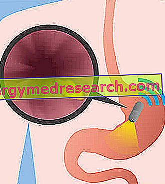
gastroscopy
Also known as esophagus-gastroduodenoscopy, gastroscopy is the endoscopy of the upper digestive tract; in other words, it is the diagnostic test that allows visual exploration from within the esophagus, stomach and duodenum.
From the executive point of view, the gastroscopy involves the use of a particular instrument, called endoscope, which the doctor gently introduces along the upper digestive tract of the patient, through the mouth. Tubular and flexible, the endoscope is equipped with a camera with a light source which, thanks to the connection with an external monitor, allows the visualization on the latter of the internal anatomy of the organs crossed. In practice, therefore, the endoscope is a probe, which the doctor inserts into the hollow organs, to study its state of health.
When the search for a stomach tumor is underway, gastroscopy is essential for the evaluation of gastric mucosa and for the identification of the possible mass of neoplastic cells.
First choice exam to detect gastric neoplasms, and in general any anomaly of the internal wall of the stomach, this instrumental test has another important value: it allows the collection of a sample of tumor cells to be subsequently analyzed in the laboratory (biopsy).
Gastroscopy requires the patient's sedation and is considered an invasive procedure.
TAC OF THE CHEST AND ABDOMEN
TAC, or computerized axial tomography, is a diagnostic test that uses ionizing radiation to create extremely detailed three-dimensional images of a more or less extensive anatomical area of the human body.
With reference to the thorax and abdomen, the TAC allows the vision of the thoracic and abdominal organs, and the detection of any anatomical anomalies or pathologies against them.
In the case of a stomach tumor, the CT scan of the chest and abdomen allows us to highlight various features of the neoplasm, including:
- The position;
- The greatness;
- Relations with neighboring anatomical structures;
- The possible dissemination of metastases in the perigastric lymph nodes, in the neighboring organs (eg: liver) and in the lungs.
Exposure of the patient to a non-negligible dose of ionizing radiation makes the CT scan of the chest and abdomen (as well as all other types of CT scans) an invasive examination.
ENDOSCOPIC ECOGRAPHY
Endoscopic ultrasound is the diagnostic test that combines the advantages of ultrasound (ie the absence of harmful radiation) with the advantages of endoscopy (observation of the organs of the human body from the inside).
In essence, therefore, endoscopic ultrasound involves the use of an endoscope, equipped with an ultrasound probe similar to those of normal ultrasound scans, and its insertion into the human body through the mouth.
The end point of the endoscope, inside the human body, is the stomach: from here the radiologist doctor collects images related to the internal gastric wall and to the neighboring organs (pancreas, perigastric lymph nodes etc.).
Endoscopic ultrasound requires the administration of a sedative to the patient and generally lasts between 30 and 60 minutes.
After its realization, the patient must wait a few hours in the hospital, so that the effects of sedation disappear; it is a purely precautionary measure.
EXPLORATIVE LAPAROSCOPY
Exploratory laparoscopy is laparoscopy with diagnostic purposes.
During his execution, the operating physician practices no more than 3 small incisions on the abdomen and, through these, introduces the laparoscope - an instrument equipped with a video camera and light source - which enables him to analyze the state of health of the abdominal organs and pelvic.
In the presence of a stomach tumor, exploratory laparoscopy is useful for studying the characteristics of the tumor mass and for the precise analysis of its dissemination in neighboring organs and lymph nodes.
Tumor biopsy
The tumor biopsy consists in the collection and histological analysis, in the laboratory, of a sample of cells from a tumor mass.
It is the most suitable test to define the main characteristics of tumors, including histology, cells of origin of neoplasia and staging .
On the occasion of a biopsy on a sample of cells belonging to a gastric tumor, a pathologist and a gastroenterologist are generally involved in the histological analysis.
STADIUM OF THE ADENOCARCINOOMA OF THE STOMACH
The parameter " staging of a malignant tumor " includes all those information, collected during biopsy, which concern the size of the tumor mass, its infiltrating power and its metastasizing capacities.
For adenocarcinoma-type stomach cancer, doctors recognize the existence of 5 levels of staging (or stages), identified with the numbers from 0 to 4; stage 0 is the least serious, stage 4 is the most serious.
Below is a more precise description of each individual stage.
- Stage 0 : the tumor mass is limited to the most superficial portion of the mucous membrane of the stomach, ie the layer of cells that makes up the inner wall of the stomach.
At stage 0, adenocarcinoma of the stomach is also called " in situ " gastric adenocarcinoma.
- Stage I : the tumor mass can involve one or more of the different cellular sheets that make up the mucous membrane of the stomach (therefore epithelium, lamina propria and muscolaris mucosae ).
A minimal presence of tumor cells is possible in no more than 2 neighboring lymph nodes.
In stage I, gastric adenocarcinoma is also called " early gastric cancer ", meaning "early gastric cancer".
- Stage II : the tumor mass has penetrated beyond the mucous membrane, therefore it involves the underlying tonache (muscular tunic and serous tunic).
Contamination of neighboring lymph nodes by metastasis is possible; if present, this contamination is equal or more extensive than in stage 1.
- Stage III : the tumor mass has expanded to the point of having invaded one or more adjacent organs and the neighboring lymph nodes.
Alternatively, it may have limited expansion to the outermost stomach tissues (without having affected other organs), but having spread metastases to the lymphatic system and contaminated several lymph nodes distant from the home site.
- Stage IV : the tumor mass has invaded the neighboring organs and has spread metastasis in organs and lymph nodes distant from the place of origin.
At stage IV, adenocarcinoma-type stomach cancer is also called " advanced gastric cancer ".
Stomach tumor and stage by stage therapeutic implications | |
| Stadium | Therapy adopted |
Stage 0 | Endoscopic resection of the mucosa. Alternatively, gastrectomy (partial or total removal of the stomach) without chemotherapy or radiotherapy. |
Stage I | Gastrectomy, possibly followed by chemotherapy and / or radiotherapy. Alternatively, if surgery is not viable, only chemoradiotherapy (ie chemotherapy associated with radiotherapy). |
Stage II | Gastrectomy preceded by chemotherapy. Alternatively, if surgery is not practicable, chemoradiotherapy. |
Stage III | It may be impossible to intervene. However, if it is possible, the therapy consists of a gastrectomy preceded by chemotherapy or chemoradiotherapy. |
Stage IV | It is generally ineffective and sometimes any form of treatment is impossible. When possible, a possible therapy only serves to temporarily improve the symptomatic picture. |