Generality
The sacral loin magnetic resonance is the important diagnostic tool that allows to visualize, in a detailed way, the terminal extremity of the vertebral column (from the lumbar tract to the coccyx).
Through the sacral loin magnetic resonance, the radiologist is able to:
- formulate diagnoses of pathologies, such as for example the herniated disc, sciatica, spinal tumors or discopathies;
- tracing back to the causes of persistent pain in the lumbar area of the back;
- to investigate the severity of congenital malformations of the spine, such as spina bifida;
- etc.
Unless a contrast medium is used, the preparation for sacral loin magnetic resonance is very simple, with few constraints to follow.
Duration of about half an hour, the sacral loin magnetic resonance is an examination with some contraindications; among these, the presence, within the human body, of devices or fragments of metallic nature, such as pacemakers or splinters, deserves a special mention.
In its conventional version (ie without contrast medium), the sacral loin magnetic resonance presents minimal risks, close to zero.
Valuable results of a sacral loin magnetic resonance are available within 3-4 days.
A brief review of what MRI is
Magnetic resonance, whose full name would be nuclear magnetic resonance, is a diagnostic test that allows the visualization of the inside of the human body, without resorting to surgical incisions or ionizing radiations, but thanks to harmless magnetic fields and equally harmless radio waves .
Virtually free of side effects and with very few contraindications, magnetic resonance imaging provides clear and detailed three-dimensional images of so-called soft tissues (nerves, muscles, ligaments, fat, blood vessels etc.) and of the so-called hard tissues (bones and cartilages). This makes it a test of absolute relevance in many fields of medicine: from traumatology to oncology, passing through orthopedics, gastroenterology, cardiology, etc.
The only limit of magnetic resonance is the high cost of the equipment, necessary for the creation of magnetic fields for the observation of the human body, and the maintenance costs of the aforementioned equipment.
What is that?
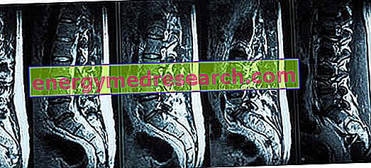
The sacral loin magnetic resonance is the nuclear magnetic resonance that allows to visualize in detail the terminal sections of the vertebral column, that is the tract of the latter which includes the 5 lumbar vertebrae, the 5 sacral vertebrae (or sacrum ) and the 4 vertebrae coccygeal (or coccyx ).
Sacral loin magnetic resonance imaging is a diagnostic test that can be performed in most hospitals and radiological clinics.
Like any other type of nuclear magnetic resonance, it is one of the practices of radiology medical specialty, so the reading of its results belongs to a radiologist doctor .
To understand: a brief review of the spine
Located in the back of the trunk, the spine or spine is the supporting bone structure of the human body.
Composed of 33-34 irregular bones, called vertebrae, the spine develops just below the skull, at the base of the back.
According to the most classic of subdivisions of the spine, the latter would include 5 sections that, starting from the top (ie from the skull), are:
- The cervical section, formed by the so-called 7 cervical vertebrae;
- The thoracic section, consisting of the so-called 12 thoracic vertebrae;
- The lumbar section, formed by the 5 lumbar vertebrae;
- The sacral section, composed of the 5 sacral vertebrae;
- The coccygeal section, composed of the 4 coccygeal vertebrae.
uses
Thanks to the sacral loin magnetic resonance, the doctors obtain clear and detailed images of the vertebrae of the tract considered, and of all that resides inside or in contact with these vertebrae, namely: spinal cord, spinal roots, intervertebral discs, pulpy nuclei, joints intervertebral, etc.
The sacral loin magnetic resonance finds employment, above all, in diagnostic field . In fact, it allows:
- Go back to the origin of a pain in the lower portion of the back, associated with an abnormal febrile state;
- Investigate circumstances such as congenital defects in the spine (eg: spina bifida );
- Analyze the precise consequences of a trauma or injury to the lower spine;
- Identify the causes of a chronic and persistent painful sensation in the lower back ( lumbago );
- Deepen a diagnosis of multiple sclerosis ;
- To confirm or not the presence of a suspected herniated disc, a discopathy or a form of arthritis in the terminal part of the vertebral column;
- Determine whether bladder and / or intestinal continence problems are due to a malfunction of the spinal nerves;
- Diagnose spinal tumors along the terminal tract of the spine;
- Diagnosing medical conditions, such as acquired syringomyelia of the lumbar sacral tract;
- Go back to the origin of leg symptoms, such as muscle weakness, numbness, tingling and lack of motor control, associated with low back pain. This symptomatology is typical of disorders such as sciatica and lumbosciatalgia .
Other purposes: intraoperative purpose
Besides being a valid tool for the identification and study of pathologies and symptoms, low-back magnetic resonance is also an intraoperative examination .
By intraoperative examination is meant a diagnostic examination carried out a few moments before a surgical operation (many times it takes place in the operating room), to observe the organ and the tissues that will be the subject of intervention a few minutes later.
Intraoperative sacral loin magnetic resonance is a practice performed very frequently just before a delicate operation on the terminal tract of the spine.
For even more precise images: sacral loin magnetic resonance with contrast
There is the possibility of increasing the sensitivity and specificity characteristics of the lumbar sacral magnetic resonance, through the use of a contrast medium, injected into the patient intravenously.
The sacral loin magnetic resonance which involves the use of a contrast medium is an example of contrast magnetic resonance .
In general, magnetic resonance tests with contrast provide three-dimensional images of organs and tissues that are more detailed and rich in details, compared to conventional magnetic resonance imaging (ie without contrast media ).
Preparation
The preparation for sacral loin magnetic resonance is the same preparation that is followed on the occasion of any conventional magnetic resonance.
This means that:
- No fasting or observance of special diets is foreseen, unless otherwise indicated by the radiologist;
- Shortly before the examination, the patient must take off any garment or object containing metal parts (bags, wallets, shoes, jewelry, etc.), answer a specific questionnaire that checks whether or not there are contraindications to the examination and, finally, communicate if you suffer from claustrophobia and, in the case of a female patient, if you are pregnant (or suspected).
Preparation for sacral loin magnetic resonance with contrast
If the sacral loin magnetic resonance involves the use of the contrast medium, the preparation for the exam varies from what was previously stated.
In fact, unlike what happens during a conventional sacral sacral magnetic resonance, the patient must:
- Complete the questionnaire that checks for any contraindications before the exam and with the signature of your primary care physician, and take it with you on the day of the procedure;
- Perform a blood creatinine test ( creatininemia ) near the MRI with contrast (not before 30 days) and take the results with you on the day of the exam, to show them to the radiologist together with the questionnaire concerning the contraindications;
- Observe a complete fast in the last 6 hours before the exam.
Procedure
The apparatus most commonly used for the sacral loin magnetic resonance is the classic machinery for nuclear magnetic resonance: a large and deep donut, capable of accommodating inside its central opening a suitably lying individual on a suitable sliding bed.
Coming to the actual procedure, this begins with the request to the patient, by a radiologist technician, to deprive himself of every forbidden object and garment, and with the answer to the questionnaire concerning the contraindications.
Once this very first part is completed, the patient must sit down on the aforementioned sliding bed, which at this time of the examination is still outside the donut machine.
In sitting on the couch, the patient receives the help of the (usual) radiologist, who, in the meantime or immediately afterwards, also takes care of providing him with all the necessary comforts (eg: pillows, blankets, earplugs, headphones etc.) and to give him the last fundamental instructions for the correct conduct of the exam. Among these indispensable instructions, the absolute immobility to which the patient must adhere during the entire procedure deserves a mention: the body's movements, in fact, jeopardize the accuracy of the images, therefore the successful outcome of the lumbar sacral magnetic resonance.
At the end of the positioning of the patient, the introduction of the couch can finally take place, in the central opening of the machinery, and the lighting of the latter (with consequent creation of magnetic fields and radio waves).
It should be remembered that modern magnetic resonance equipment is equipped with loudspeakers and cameras for communicating with medical personnel, who, as a rule, once the exam has begun, will take place in a room adjacent to where the patient resides. The presence of a communication system guarantees complete control of the situation and the possibility, to those who are submitting to the procedure, to report any problems or problems.
Like any type of MRI, even the sacral loin magnetic resonance is noisy. This explains why, before switching on the device, the patient receives the aforementioned ear plugs or the aforementioned headphones.

How does the sacral loin magnetic resonance with contrast medium vary in the procedure?
In addition to what has been reported previously, the sacral loin magnetic resonance with contrast medium includes injection of the contrast medium. This fundamental passage takes place shortly after the patient is settled, but a few minutes before the diagnostic machine is turned on.
To perform the injection is the radiologist doctor, in collaboration with a professional nurse.
Generally, the contrast medium used is an aqueous solution containing a certain percentage of gadolinium (a gadolinium -based contrast agent).
How long does a luminous sacral magnetic resonance last?
Usually, the sacral loin magnetic resonance examinations last approximately 20-30 minutes .
The possible use of the contrast medium increases the aforementioned time by a few minutes.
What happens at the end of the exam?
Generally, immediately after conventional sacral loin magnetic resonance, the patient can get dressed and return home and to his daily activities, waiting for the medical response.
In the event that the contrast medium has been used, the return home can take place only 1-2 hours after the end of the procedure. This is a precautionary measure, adopted due to the possible adverse effects of the contrast agent.
risks
Conventional sacral loin magnetic resonance is a highly safe and completely harmless instrument for the human body.
The advantage of not exposing the patient to ionizing radiation makes it a test that can be repeated several times, even after a short time.
The risks of sacral loin magnetic resonance with contrast
Although very rarely, contrast magnetic resonance procedures - including those that have as their object the terminal tract of the spine - can give rise to adverse effects of varying severity.
The mildest adverse effects include: headache, nausea, vomiting and / or dizziness.
The most serious adverse effects, on the other hand, include: allergic reactions to the contrast medium and systemic nephrogenic fibrosis (*).
Contraindications
Conventional sacral loin magnetic resonance has the same contraindications as all other types of magnetic resonance imaging that do not use contrast media.
Consequently, all those who present, within the human body, devices or fragments of metallic nature, such as for example pacemakers, neurostimulators, splinters (those in the eye in particular) and intracranial clips, cannot undergo the diagnostic examination in question. for a brain aneurysm, hearing aids, metal prostheses, metal sutures, etc.
MRI and pregnancy
For pregnant women, the examination is not recommended during the first 3-4 months of pregnancy; after which there is more freedom.
Contraindications of magnetic resonance sacral resonance with contrast medium
Briefly, in the case of a lumbar sacral magnetic resonance with contrast, the following contraindications are added:
- The presence of severe renal insufficiency or abnormal creatinine test results ;
- The presence of severe liver failure ;
- The presence of an allergy against the contrast medium necessary for the examination;
- The state of pregnancy .
Results
As a rule, the results of a lumbar sacral magnetic resonance with diagnostic purposes are available to patients 3-4 days after the examination.
Like any other type of magnetic resonance, sacral loin magnetic resonance can provide fundamental elements for a precise diagnosis.