Generality
Carotid stenosis is a disease that affects the carotid arterial system.
The term stenosis, in this case, indicates a reduction in the vessel size, due to which the blood flow downstream of the narrowing is decreased; it follows a state of suffering of the organs reached by it, due to lack of oxygen and nutrients carried by the blood.

The main cause of carotid stenosis is atherosclerosis, a particular form of arteriosclerosis that affects the large vessels.
What is carotid stenosis. Pathological anatomy
The stenosis (from the Greek στενόω ‰, narrow) of the carotid is the narrowing of the carotid vasal lumen. Before proceeding with the detailed description of the pathology, it is useful to briefly recall the anatomy of the carotid system. The latter is composed of:
- Two common carotid arteries, right and left.
- Two branches for a single common carotid artery: the internal and external carotid artery .
- Collateral branches, which arise from the internal and external carotids.
The carotid system, through its various ramifications, goes to flush the cerebral districts and the areas of the head corresponding to the face and eyes. The partial or total occlusion of the carotid artery results in an ischemic type phenomenon affecting the perfused tissues, since the flow rate of the blood pumped by the heart is compromised. The outcome of a carotid occlusion is clearly dramatic, as non-oxygenated tissues undergo necrosis (cell death). Necrosis of the tissues can be followed by a stroke and, when carotid stenosis is severe, the death of the sick individual.
From the studies of pathological anatomy (that is, on how a tissue or organ affected by a pathology appears), conducted on the carotids affected by stenosis, the following characteristics emerged:
- Occlusion is more frequent in the left carotid artery, which arises directly from the aortic arch, in the thorax. The reason is the following. Atherosclerosis preferably affects the large vessels, and, in the case of the left carotid artery, the direct connection with a larger vessel predisposes the vessel to risks of stenosis, of atheromatous origin, higher than the right carotid artery; the latter, in fact, is born from the anonymous artery, which in turn originates from the aortic arch.
- Brain injuries, due to ischemia, are more or less marked based on the extent of carotid narrowing. There is a direct proportionality: a greater occlusion of the vessel, therefore, means a more serious damage and a progressive worsening of the symptoms.
- Occlusions usually occur at the level of the bifurcations and at the origin of the collateral branches of the carotids.
Carotid stenosis is a typically male pathology, since atherosclerosis, the main cause of stenosis, affects men more than women. Moreover, it is a pathology that does not spare anyone, since atherosclerosis is a condition that, sooner or later, afflicts every individual.
Causes of carotid stenosis. Pathophysiology
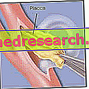
The main cause of carotid stenosis is atherosclerosis, a particular form of arteriosclerosis, which affects, preferably, large-caliber arterial vessels. Atherosclerosis is characterized by the appearance, at the level of the intimate habit and the innermost layers of the middle layer of the arterial vessel, of a detected plaque with precise contours. This focus is called an atheroma . The atheroma has a fibrolipid consistency: the fibrous component is due to a proliferation of the fibrous connective tissue ("cicatricial" tissue); the lipid component instead comes from the blood plasma and consists of cholesterol crystals, triglycerides and fatty acids.
The onset of an atheroma is due to several factors, all of which are equally important. The best known are:
- Hypertension
- Obesity
- Smoke
- Hypercholesterolemia
- Sedentary life
- Diabetes
- Aging
The atheroma, which develops at the level of the vessel's intimate habit, arises following an imbalance between the vessel wall and blood circulating in the lumen of the artery. In other words, the factors that induce atherosclerosis cause the blood flow in the vessel to be altered to such an extent as to generate a lesion in the vessel wall, ie in the endothelium. The lesion creates an inflammatory situation and draws blood plasma cells, such as red blood cells and white blood cells, whose intervention generates the first small plaque. High blood pressure, for example, creates a swirling flow inside the arteries. This explains why the atheromas develop electively where there are carotid bifurcations: here the stresses to which the vessel is subjected are superior. Another example of instability in the relationship between the inner wall of the carotid artery and the blood concerns aging, an event that affects every individual. It reduces the elasticity and contractility of the arteries, thus modifying the blood flow.
The picture is also enriched with the formation, at the atheroma level, of a thrombus . Thrombus is a solid mass of blood cells. The consequence is natural, because, where a lesion is created, there is also a recall of platelets, or thrombocytes, and factors that deal with the coagulation process. These actors combine to increase the thickening of the atheroma. At this point, the lumen of the arterial vessel of the carotid artery narrows further.
To make the situation even worse, it is the possibility that the thrombus flakes off into smaller particles, which are lost in the bloodstream. These free particles, called emboli, can reach the brain, accelerating the processes of cerebral ischemia and stroke .
Other causes of carotid stenosis are:
- Aneurysms
- Fibromuscular dysplasias
- arteritis
- kinking
- Coiling
Symptoms and signs
A clinical sign of a carotid stenosis is the absence of pulsations in the affected vessel. The verification is carried out by palpation and has a certain degree of uncertainty. In fact, pulsation may also be present in conjunction with a narrowing of the carotid artery.
The main sign that characterizes a carotid stenosis is the so-called transient ischemic attack, also known as TIA . It is defined as transitory, since it has a duration limit: no more than 24 hours. The ischemic attack occurs at the cerebral, facial and ocular levels, ie the areas not sufficiently supplied by the occluded carotid artery. The clinical signs, due to the TIA, manifest themselves with:
- Loss of limb control: hemiplegia of the side opposite to that of the occluded carotid artery. This is explained because - for example - the right hemisphere of the brain, sprayed by the right carotid artery, controls the limbs of the left side of the body.
- Difficulty in speaking : the language sometimes becomes incomprehensible.
- Eyesight problems : split or blurred vision. Possible blindness, which occurs initially with a black or gray veil that falls in front of the eye. In this case, the affected eye is on the same side as the occluded carotid artery.
- Failure to coordinate walking.
- Paresis of the face.
If the stenosis involves ischemic damage of greater magnitude, which lasts up to 3 days, we speak of RIND, that is reversible ischemic neurological deficits . The symptoms are similar to those of TIA.
Finally, if the occlusion of the carotid artery is severe and almost, if at all, complete, the resulting symptom is ischemic stroke, or stroke . The consequences are obvious and no longer transitory: the individual, who is affected by it, completely loses the sensitivity, the faculty of movement and the different functions controlled by the areas no longer oxygenated by the blood stream. In most cases, this situation leads to death.
Diagnosis
A first diagnosis of carotid stenosis can be based on the monitoring, by simple palpation, of the carotid pulse. The absence of pulsation at one of the two carotids may mean that an occlusion exists.
An important test is the so-called carotid artery sign, useful for determining not only the presence of stenoses, but also which of the two carotid pathways is occluded. It consists of alternately compressing one of the two carotids, interrupting the blood flow that flows through the carotid vessel. If the compressed carotid is the healthy one, after a time varying from 10 to 30 seconds, the patient shows signs of malaise, pallor and loss of consciousness. If the compressed carotid artery is the already occluded one, the patient does not manifest symptoms, since the opposite way, patent, compensates for the lower inflow, due to the stenosis, to the cerebral districts.
Instrumental diagnostic tests consist of:
- ecodoppler
- Digital angiography
- Angioscanner
- Angio
Ecodoppler . This is a non-invasive test, useful for the doctor to identify the position of the atheromatous plaque and the degree of stenosis, ie when the lumen has shrunk. In fact, it is a method that allows, through an ultrasound scan, to observe the morphology of the vessel walls and identify any anomalies; through a doppler, on the other hand, it is possible to evaluate, with an ultrasound analysis, the hemodynamic situation, ie the speed of the blood flow, in the area of the carotid artery affected by the plaque. This last datum, that is to what the blood travels in the point of occlusion, reveals the degree of stenosis of the atheromatous plaque.
Digital angiography . It is the most accurate survey and is useful for assessing the degree of stenosis. It consists in injecting an iodinated contrast agent into the arterial circulation, by means of a catheter. The catheter is conducted into the area to be investigated. In this area, the catheter path is followed by radiographic equipment, which shows the internal structure of the carotid artery.
Computed tomographic angiography, or CT angiography . It is based on the scanning of the carotid area. The images, obtained by radiographic instrumentation, show the three-dimensional structure of the carotid vasal cavities. Requires injection of an iodinated contrast agent.
Angio-magnetic resonance imaging, or angiography . The examination uses a paramagnetic contrast agent, which is injected into the patient. It allows to assess the site and extent of carotid vasal lumen alterations.
Therapy
The pharmacological therapy is useful to improve the patient's symptoms or to prevent their deterioration, but does not "adjust" a lesion, such as the atheroma, present on the arteries. It provides for the administration of:
- Blood-thinning drugs . They are used to avoid the formation, or the worsening, of the thrombi present in the areas affected by the atheromas. The worsening of a thrombus can degenerate, as previously said, into an embolus. To thin the blood, the patient can be given:
- Platelet aggregation inhibitors. Platelet aggregation and lump formation decrease. One of the most used is aspirin.
- Anticoagulants. They act on coagulation factors. They should be used with caution, before surgery or if the patient is suffering from other diseases that require anticoagulant treatment. One of the most used is the coumadin.
- Drugs that limit the evolution of atheromatous plaque
- The lipid-lowering agents. The rate of cholesterol and triglycerides in the blood decreases, ie the lipids that act in plaque formation.
- Antidiabetics. They are indicated for diabetics. Diabetes is a condition that predisposes to carotid stenosis.
- Antihypertensives. They serve to normalize blood pressure. The vortex blood flow, generated by hypertension, favors the lesion of the intimate vessel of the vessels and the consequent formation of atheromatous plaques.
Surgery, on the other hand, is the only therapeutic approach useful for restoring normal blood flow within the occluded carotid artery.
Two types of intervention are possible:
- Endarterectomy . This operation eliminates the atheromatous plaque and any lumps and residues, related to thrombi and emboli, respectively.
- Angioplasty and carotid stenting . The operation serves to "repel" the atheromatous plaque, re-establishing the normal size of the carotid vessel lumen. It is practiced under local anesthesia. The vascular surgeon operates by using two catheters : one is provided with a metal mesh ( stent ) and another with a balloon . Introducing them into the arterial circulation and reaching the area affected by the atheroma, the doctor ensures that the normal diameter of the occluded carotid artery is restored by means of the balloon, and that enlargement is maintained by means of the metal mesh. The balloon is inflated only once the catheter has been conducted into the area affected by the plaque. Later it will be removed.


Surgery is necessary when carotid occlusion affects more than 70% of the vessel lumen. The same applies to cases in which, although the shrinkage is lower in terms of percentage, the symptomatology foresees the possibility of critical situations, such as TIA, RIND or stroke. In the absence of these serious symptomatic conditions and at stenosis rates lower than 70%, the intervention is not a priority. The reason is due to the extreme delicacy of surgical operations involving the carotid artery. When the patient has an advanced stage of carotid stenosis, the risks associated with the operation do not exceed those that could create a stroke. Therefore, plaque is eliminated.