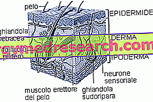
The dermis is the middle layer of the skin, between the hypodermis and the epidermis. Unlike the latter, from which it is separated from the basement membrane, the dermis is richly vascularized and innervated.
The dermis performs functions of mechanical and metabolic support towards the epidermis, to which it transfers nutrients and sebum, an oleaginous substance that protects the superficial layer of the skin from bacteria and dehydration. It has a wavy shape due to the presence of dermal papillae, extroflexions that have the purpose of inserting themselves into the ridges present in the overlying epidermal layer. This particular anatomical conformation has the purpose of increasing the adhesion between the two layers and of favoring metabolic exchanges.
From a histological point of view, the dermis is a connective tissue composed of fibrous glycoproteins immersed in a fundamental substance. Inside there are different types of cells, hair follicles and glands typical of the skin.
The dermis can be divided into two portions. The most superficial layer, called adventitial, is rich in cells; in the deeper one, called reticular, fibers prevail instead.
In the dermis three components can be distinguished: the cells, the fibers and the fundamental substance (or amorphous) that fills the spaces left free by the fibers and dermal cells.
CELLS: fibroblasts are the most abundant cells in the dermis and are responsible for the synthesis of fibers and the components of the fundamental substance.
In addition to fibroblasts, mast cells are also present, cells that contain many granules rich in heparin (anticoagulant agent) and histamine (mediator of inflammatory reactions).
The dermis is also populated by cells from the blood such as macrophages, granulocytes and lymphocytes. The presence of these cells at the level of the dermis increases during inflammatory states. In particular, macrophages derive from blood monocytes which, after leaving the capillaries, take on an appearance similar to fibroblasts and are called histiocytes. When an inflammatory process is in progress the histiocytes increase in size and acquire the ability to incorporate foreign particles and necrotic material (phagocytize). In this case the histiocytes are called macrophages, which belong to the family of antigen-presenting cells and play a leading role in the immune response.
The dermis performs important metabolic, immunological, thermoregulatory and sensitive functions, as well as support. At this level we find important structures, such as the sweat and sebaceous glands, the roots and the hair bulbs, the erector muscles of the hair and a dense network of capillaries. |
FUNDAMENTAL SUBSTANCE: it consists of glucosaminoglycans (GAG). These are polysaccharides consisting of long chains of disaccharides, in which at least one of the two units is an amino sugar (glucosamine or galactosamine).
The best known glucosamminoglycans are hyaluronic acid and heparin. These and other substances belonging to this family have the ability to retain a lot of water, forming a gel.
A gel is a state in which a dispersed phase and a dispersing phase coexist. In the specific case the molecules of glucosamminoglycans (dispersed phase) form a sort of lattice between whose meshes water (dispersing phase) is contained.
At the level of the dermis, this gel occupies most of the extracellular space and is responsible for skin turgidity . Glucosamminoglycans are rather rigid molecules that do not fold up, consequently they assume rather extended conformations (called random coils) and occupy an exaggerated volume compared to their mass.
In the dermis all the glucosamminoglycans present, except hyaluronic acid, bind in large numbers to a single filamentous protein (of the core or protein core), forming proteoglycans.
Many proteoglycans bind to one core of hyaluronic acid forming huge aggregates:

FIBERS: the main ones are those of collagen. Collagen is an extremely complex glycoprotein organized in large fibrous bundles and, in addition to being the body's most abundant protein, it alone accounts for 70% of proteins in the skin.
Collagen has a supporting function and gives considerable mechanical resistance to the dermis. In the most superficial layer, called adventitial, there are also thinner collagen fibers, called reticular ones.
In addition to collagen fibers, in the dermis there is a small proportion of elastic fibers which, together, represent only 2% of cutaneous proteins. They consist of elastin which gives the skin a certain degree of elasticity, essential both to allow facial expressions and to follow the numerous variations in body size that occur throughout life.
The elastin molecules are joined by cross bridges, thanks to which they form a wide net that gives the skin a fair degree of elasticity. However, skin distension is limited by the presence of collagen fibers mixed with elastic ones. However, there are cases in which the distension of the skin is so pronounced as to cause the breakage of the collagen fibers: a classic example is given by gravid stretch marks.
Ipoderma »