Locomotion is the ability of animal organisms to move, moving from place to place.
The movement is made possible by the anatomical conformation of the skeleton that is set in motion by muscle contraction. Other anatomical structures (tendons, nerves, ligaments, etc.) participate in the movement, forming together the so-called locomotor apparatus.
The locomotor apparatus is in turn composed of three distinct apparatuses or systems:
- skeletal system constitutes support and insertion for muscles and protection for internal organs. It is PASSIVE system in movement: it is the skeletal segments that are moved following the muscular action.
- articular apparatus constituted by the regions in which the bone segments face with the relative annexes
- muscular system. It is the active element in locomotor mobilization and gives the movement of the viscera and their portions.
Each of these systems can suffer more or less serious injuries during the lifetime and, while some heal spontaneously, others require surgery and / or pharmacology.
The most frequent traumatisms of the locomotor apparatus are listed below.
Injuries of the bone apparatus
1) FRACTURES: fracture means an interruption of the structural integrity of a bone.
Traumatic fractures are distinguished, in which the trauma acts on a normal bone, and pathological or spontaneous fractures, which are produced by weak traumas capable of overcoming the resistance of an altered bone, but not that of a normal bone. Fractures can be located exactly at the point where the causes have exerted their action (direct fractures) or, on the contrary, reside in a more or less distant point (indirect fractures). They are called complete if there are two or more distinct fragments, otherwise they are incomplete and in this case they can be exposed or covered, depending on whether or not there is a discontinuity of the overlying soft parts; finally we speak of comminuted fractures, when the bone is reduced in multiple fragments or splinters.
Therapy: must tend to obtain a bone scar called callus and can be summarized in two words: reduction, restraint.

In any case, the reduction of fractures must be practiced as soon as possible after the trauma (before too much swelling of the soft parts is produced and a re-starting of the bone ends), under radiological control, and, in general, under local or general anesthesia; this suppresses the pain felt by the injured person when the bone fragments are mobilized and decreases the resistance opposed to the surgeon's effort by the contraction of the muscles that approach the fracture.
After reducing the fragments it is necessary to maintain the reduction: this is done with the help of some devices that ensure the absolute immobility of the fragments; the simplest appliances are formed by flexible and resistant sheets (splints) that are applied along the fractured limb to keep it immobile: these splints are generally made of wood or flexible or rigid metal, circular bandages will be avoided, since the tissues, tumefacing, they would be too compressed. In urgent cases the slats can be replaced with pieces of wood.
In short, it is a question of improvising two lateral, wide defenses, which rise up the entire length of the limb, in order to immobilize the two joints above and below the fractured part.
Cushions can also be used to isolate the splints from the limb and bandages surrounding the limb, designed to keep the different parts in contact and to form a whole. Several mobile bandages have been reported, which use these elements: the main ones are the spiral bandage, the scultet apparatus. If nothing can be found that can be used, the resource of attaching the injured lower limb to the healthy limb always remains, the latter taking the place of a splint, for the arm, the side of the chest will serve as defense.
Whenever possible, the immovable ones, which are generally made of plaster, are preferred to fixed appliances.
The plaster is cut according to the region to be cast in circular bandages, in strips or in showers. These gypsum appliances must be monitored during the days following the application, as they can be too narrow and cause local compression, very frequent at the heel and malleoli. They can also give rise to pain and be the cause of eschar. Sometimes instead they become too loose, when the limb deflates and loses weight; the displacement is then produced, hence the need to redo a new appliance.
The duration of the application of these devices is variable for each fracture.
The harassment of all these devices, mobile or immovable, is that they soon find ankylosis and muscular atrophy. To avoid them you need to immobilize your joints as little as possible and then use massages and electricity.
At present, fractures are increasingly being treated surgically, in which the plaster realizes insufficient immobilization of the fragments. The main procedures adopted are bone suture, osteosynthesis, incarceration and in the most serious cases nailing is used.
The latter also called screwing is a method that makes it possible to make two fragments of spongy bone solid in whose cavity a nail is fixed which unites the two parts of divided bone.

3) COMMITMENT: shock produced in the body by a fall or a violent collision, therefore two types of commotion can be distinguished:
"electric commotion" when there is a contraction caused by an electric current and "concussion" when there is a loss of knowledge, generally transient and reversible, that does not produce permanent damage but can degenerate into a coma.
Cranial trumatism always exposes the risk of injuring the brain more or less severely. Therefore, in the hours following the trauma, signs of a cerebral contusion, a hematoma and other more or less serious characteristics that require more detailed examinations and a surgical operation can be observed.
Injuries of the muscular system
CONTRACTURE: Continuous and involuntary contraction of one or more muscles, whose rigidity is such as to form hard, appreciable cords under the skin. When it hits a limb, it immobilizes it in a more or less strong flexion or extension; to the face, it does not allow to open the jaw. The contracture can occur suddenly or follow convulsions or muscle paralysis. It ceases under the action of chloroform, which distinguishes it from muscular retraction, in which there is the alteration of the muscle fibers, while in the contracture there is simply an exaggeration of the function. The contracture is often painful.
CONTUSION: lesion produced by a shock, without the continuous solution of the skin and with transfer of blood.

STRAPPO: partial or total laceration of a muscle's fibers, following a violent movement.
STRETCH: excessive elongation, beyond the physiological threshold, of the muscle fibers.
Injuries of the joint apparatus:
DISTORTION: Traumatism of a joint, due to a forced movement and which is accompanied by elongation or rupture of the articular ligaments, without following a permanent displacement of the articular extremities. It is the first stage of a dislocation or, if you want, a missed dislocation. Distortion is characterized by ligament injuries, lesions of the joint capsule and the synovium, and especially vasomotor disorders; lively pain, local heat, swelling (bruising) and considerable hydrartre.

Therapy: in sprains without serious ligament injuries, local infiltration of novocaine was recommended, which eliminates pain and vasomotor disorders and allows immediate use of the limb. The massage, followed by a bandage, is proposed for the same purpose. If there are ligamentous lesions, we must not resume walking, but immobilize with plaster. Physiotherapy, mineral water treatments can be used to combat after-effects.
LUXURY: permanent displacement of two articular surfaces, due to external violence, or alteration of the tissue of one of the parts of the joint. Depending on whether the relationship between the joint surfaces is completely or partially suppressed, the dislocation can be complete or incomplete (sub dislocation). Sometimes the lesion is limited to an opening of the joint capsule and to the partial rupture of the ligaments, but often these are torn and can also remove bone fragments; the muscles are violently bruised; a blood effusion is formed. In general, everything comes back to place after the dislocation is reduced.
Symptoms: pain on a very large surface, exacerbated by movement, attenuated by immobility; deformation, particular attitude of the limb, whose length is modified (shortening or lengthening); abolition of active movements while some passive movements remain (exaggeration of the abnormal limb situation) and abnormal movements.

Therapy: do not try to reduce the dislocation, since it is a delicate maneuver that only a doctor will be able to do. By trying to reduce dislocation, you risk tearing vessels and nerves and causing a fracture. For the reduction the doctor uses, depending on the case and according to whether the dislocation is more or less recent: o sweetness maneuvers, which consist in methodically pressing on the displaced part, so as to push it towards the normal articular cavity, or force maneuvers . With the latter the body is held firmly still (against extension), then a traction effort is made on the dislocated limb (extension), either directly or by means of an elastic strap. The reduction then occurs naturally or with a surgical procedure. Anesthesia helps overcome muscular endurance. In cases of irreducible dislocation (due to the interposition of muscular or tendinous parts between the articular surfaces) or long-standing dislocations with adhesions, it is necessary to resort to surgery (bloody reduction). After the reduction the immobilization is necessary for a variable period of time.
Paramorphism: acquired alteration of the external form of the body and its usual functional attitudes, due to asthenia and hypotonia of the muscles and ligaments.
PARAMORPHISMS OF THE VERTEBRAL COLUMN:
SCOLIOSIS: scoliosis involves a lateral shift of the spine
CYCLOS: the kyphosis involves an exaggerated dorsal arching
LORDOSI: in lordosis there is an accentuation of the lumbar curvature
In all three cases mentioned, it is necessary to intervene early with gymnastics and, possibly, with special corsets to prevent the malformation from becoming definitive. Very important, for the purpose of a harmonious development of the entire skeletal framework, is also the control of the structure of the foot which, being the "base" of support of the body, directly influences the conformation and the arrangement of the supporting bone elements. In the normal foot, the weight of the body is supported in the plantar arch. However, the case in which the plantar arch is not well conformed may occur and in this case the situation of "flat foot" occurs. To avoid this defect, a correct ambulation setting is required, but above all a careful choice of footwear. Shoes that are too narrow on the toe or with exaggeratedly high heels force the feet to take a forced position by compressing or deforming them. Therefore, heels are recommended not higher than 2cm for children, and not more than 6cm for adults, and possibly the presence of orthotics that keep the arch sufficiently raised.
PARAMORPHISMS OF THE FOOT:
FLAT FOOT (described above)
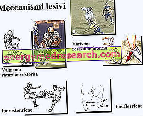
VARISM: position anomaly whereby the longitudinal axes of two contiguous skeletal segments or of two parts of the same segment do not coincide on the frontal plane (imaginary plane passing tangentially to the forehead), but they form an open angle inside with respect to the midline of the body. The opposite anomaly is valgism.
VALGISMO: defective attitude of two contiguous skeletal segments (or of two parts of the same segment) so that their longitudinal axes do not coincide on the frontal plane (imaginary plane tangential to the front), but they form an angle open towards the outside (with respect to the line median of the body). The opposite anomaly is varus. The causes of valgus are various: congenital malformations, rickets, polyomelitic paralysis, traumatisms. Particularly important are valgus of the knee (valgus knee) and femoral neck (coxa valga).
PARAMORPHISMS OF THE KNEE:
1) VARISM (see varus in the foot parameters)
2) VALGISMO: (see valgism in the foot parameters).