Generality
Syndesmosis is a particular type of fibrous joint (or synarthrosis).
The peculiarity of this type of articulation lies in the fact that it unites two bones by means of an aponeurotic membrane or by means of a series of ligaments.

In the human body, the most important examples of syndesmosis are: tibio-fibular syndesmosis, located between the tibia and the fibula, the radio-ulnar syndesmosis, located between the radius and ulna, and the vertebral syndesmosis, located in the vertebral column.
Syndesmosis are fibrous joints that can be injured. In general, these are injuries of a traumatic nature.
Short anatomical reference on the joints
The joints are anatomical structures, sometimes complex, which put two or more bones in mutual contact. In the human body, there are about 360 of them and their task is to keep the various bone segments together, so that the skeleton can fulfill its function of support, mobility and protection.
The anatomists divide the joints into three main categories:
- Fibrous joints (or synarthrosis ). They generally lack mobility and the constituent bones are held together by fibrous tissue. Typical examples of synarthrosis are the bones of the skull.
- Cartilaginous joints (or amphiarthrosis ). They have poor mobility and the constituent bones are joined by cartilage. Classic examples of amphiartrosis are vertebral vertebrae.
- Synovial joints (or diarthrosis ). They are highly mobile and include various components, including: the articular surfaces and the cartilage that covers them, the joint capsule, the synovial membrane, the synovial bags and a series of ligaments and tendons.
Typical examples of diarthrosis are the joints of the shoulder, knee, hip, ankle, etc.
What is a syndesmosis?
A syndesmosis is a particular type of fibrous joint that unites two bones separated by a discrete space, through a sort of membrane and / or a network of ligaments .
The membrane and ligaments responsible for connecting the bones forming syndesmosis are named, respectively, of interosseous membrane and interosseous ligaments .
The synesmosis of the human body - to be precise some synesmosis of the human body - are rare examples of synarthrosis that can enjoy minimal mobility.
Features
Syndesmosis lacks a joint cavity and lining cartilage.
The interosseous membranes and the interosseous ligaments, of which they can be equipped, have the task of containing and guaranteeing stability to the bone components involved in the joint.
Specifically, interosseous membranes are aponeurotic membranes . Also known as aponeurosis, an aponeurotic membrane is a thin lamina of fibrous tissue, similar to a tendon, with a shiny white-silver appearance.
Interosseous ligaments, on the other hand, are very resistant bands or bundles of fibrous connective tissue.
Examples
The most important synesmosis of the human body is tibio-fibular syndesmosis, located between the tibia and fibula (or fibula), the radio-ulnar syndesmosis, located between the radius and the ulna, and the syndesmosis of the vertebral column.
TIBIO-FIBULAR SYNDESMOSIS
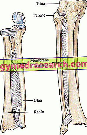
Tibio-fibular syndesmosis, or tibio-peroneal syndesmosis, includes an interosseous membrane and a network of interosseous ligaments, which develop from the lateral portion of the tibia to the medial portion of the fibula .
Tibia and fibula are the two bones constituting the skeleton of the leg .
Equipped with small holes for the passage of vessels and nerves, the interosseous membrane involves, in full, the bodies of tibia and fibula (NB: the bodies are the central bony portions). The interosseous ligaments, on the other hand, affect the distal portions of the tibia and fibula; these ligaments are altogether four and are better known as: anterior inferior tibio-fibular ligament (or anterior distal tibio-fibular ligament), inferior posterior tibio-fibular ligament (or distal posterior tibio-fibular ligament), transverse ligament and interosseous ligament.
The functions of tibio-fibular syndesmosis are at least four:
- Keep the tibia and fibula together during the movements of the lower limbs, especially the leg;
- Inserting to the leg muscles known as: extensor muscle along the toes, extensor muscle of the big toe, tibialis anterior muscle, tibialis posterior muscle and anterior peroneal muscle;
- Transfer to the fibula some of the weight-forces that weigh on the tibia;
- Provide stability to the ankle joint.
Some anatomy experts believe that the interosseous membrane, interposed between tibia and fibula, constitutes a syndesmosis and that the interosseous ligaments, interposed between the distal portion of the tibia and the distal portion of the fibula, form another syndesmosis.
In other words, they divide the so-called tibio-fibular syndesmosis into two distinct entities, one that includes only the interosseous membrane and one that includes only the interosseous ligaments.
RADIO-ULNAR SYNDESMOSIS
Radioulnar syndesmosis includes an interosseous membrane, which develops from the medial portion of the entire radium body to the lateral portion of the entire ulna body. Radio and ulna are the two bones making up the skeleton of the forearm .
Just like the interosseous membrane of the tibio-fibular syndesmosis, the interosseous membrane of radioulnar syndesmosis has small holes through which blood vessels and nerves pass.
There are at least four functions of radioulnar syndesmosis:
- Keep together radius and ulna, during pronation and supination movements of the forearm;
- Insert some muscles of the anterior and posterior compartments of the forearm;
- Transfer to the ulna part of the weight-forces that weigh on the radio;
- Provide stability to the wrist joint.

SYNDESMOSIS OF THE VERTEBRAL COLUMN
Introduction: to understand the location of the symphysis of the vertebral bodies, it is essential to briefly review the anatomy of the vertebral column and vertebrae.
Supporting axis of the human body, the vertebral column or spine is a bone structure of about 70 centimeters (in the adult human being), which includes 33-34 vertebrae stacked on each other.
The vertebrae of the spine have a general structure quite similar to each other. In fact, they all have:
- A body, in anterior position
- A bow similar to a horseshoe, in a rear position.
A generic vertebral arch includes: two peduncles, two transverse processes, two upper joint processes, two lower joint processes, a spinous process and two laminae.
- A vertebral hole, deriving from the particular arrangement of the arch with respect to the body.
The syndesmosis of the vertebral column are those fibrous joints that unite the spinous processes and the laminae of two adjacent vertebrae.
Formed by interosseous ligaments, the syndesmosis of the vertebral column represent a singular case of fibrous articulations endowed, albeit minimally, with mobility.
diseases
Syndesmosis can be the subject of injuries, in which the interosseous ligaments stretch too much or the interosseous membrane suffers an injury.

Figure: reproduction of a generic vertebra of the human body. In red, a vertebral lamina is highlighted. Image from the site: lancsteachinghospitals.nhs.uk
In general, injuries to syndesmosis depend on traumatic bone fractures.
For example, injuries to tibio-fibular syndesmosis often result from fractures or sprains of the ankle, resulting from sports injuries or road accidents.
To diagnose an injury to a syndesmosis and to quantify its severity, the following are fundamental: the physical examination, the anamnesis and the images provided by X-rays, a CT scan or an MRI scan.
Depending on the severity of the accident that has affected a syndesmosis, the treatment can be conservative or surgical: in general, it is conservative if the syndesmosis has suffered a slight injury, while it is surgical if the syndesmosis has been the subject of a severe injury.


