Generality
The muscles of the leg are the muscles with total or partial seat in the skeletal compartment between the thigh and the foot and which includes the tibia and fibula bones.

Leg muscles contribute to fundamental movements for locomotion. In this case, they contribute to: plantarflexion, dorsiflexion, extension of the leg and toes, flexion of the leg and toes, eversion of the foot and inversion of the foot.
Brief anatomical reference
The muscles of the human body have two ends: an initial or proximal call and one called terminal or distal.
At each end, there is a tendon. Tendons are formations of fibrous connective tissue that join two distinct bones or two different parts of the same bone.
Then, the muscles find insertion on the skeleton, by means of the tendons.
The anatomical texts and experts have a tendency to identify the initial extremity and the terminal extremity of a muscle with the tendon present on each of these extremities.
Anatomical meaning of proximal and distal
Proximal and distal are two terms with opposite meaning.
Proximal means "closer to the center of the body" or "closer to the point of origin". Referring to the femur, for example, it indicates the portion of this bone closest to the trunk.
Distal, on the other hand, means "farther from the center of the body" or "farther from the point of origin". Referred (always to the femur), for example, it indicates the portion of this bone furthest from the trunk (and closer to the knee joint).
Definition
The muscles of the leg are the muscles whose fibers reside totally or only partially in the anatomic-skeletal section constituted by the tibia and the fibula.
The fact of residing completely or only partially in the leg does not necessarily mean that their extremities are attached to the skeleton of the leg.
The leg is the anatomical region of the human body between the thigh and the foot.
Both at the border between the thigh and leg and at the border between the leg and the foot, there is an articulation: in the first case, it is the knee joint; in the second case, however, it is the ankle joint.
TIBIA AND PERONE: A BIT OF ANATOMY
Tibia and fibula are the two equal, longitudinal and parallel bones that make up the skeleton of each leg.
Both belonging to the category of long bones, they adjoin the femur, superiorly, and the astragalus, inferiorly. Femur and astragalus are, respectively, the only bone that constitutes the skeleton of the thigh and one of the 7 tarsal bones of the foot.
Tibia and fibula have some peculiarities that, in this case, deserve a brief reminder, as they are fundamental to understand the muscular structure of the leg.
* NB: in the following text and also in the following ones, the reader will encounter the terms medial and lateral; for those who did not know the meaning, they can consult present at the end of the description of tibia and fibula.
- Tibia . Located on the inner side of the leg, in a medial position with respect to the fibula, the tibia is a long bone particularly broad at the ends and somewhat slender in the central portion.
Proximal end (or proximal epiphysis): it is the bone portion on the border with the femur. It has some very important anatomical structures, such as the two condyles (medial and lateral), the tibial plateau and the tibial tuberosity.
From the functional point of view, it plays a leading role in the knee joint (NB: it articulates with the condyles of the femur) and in the upper tibio-fibular joint. The upper tibio-fibular joint is the proximal junction point between tibia and fibula.
Body (or diaphysis): is the central bony portion, between the proximal end and the distal end. It has three surfaces: a medial, a lateral and a posterior.
On the lateral surface takes place the so-called interosseous membrane, which, by binding to the fibula, forms a fibrous articulation.
Distal end (or distal epiphysis): it is the bony portion on the border with the talus of the foot. At the lower edge, it has a cavity (the mortar) that serves to form the ankle joint. On the inner side (in medial position), it presents a bone process, called tibial malleolus (or medial malleolus); the purpose of the tibial malleolus is to give stability to the ankle. Finally, on the external side (in medial position), it has an articular facet (the so-called fibular incisura), which, joining the fibula, forms the inferior tibio-fibular articulation.
Functions: the tibia has the task of supporting the weight of the body, absorbing part of it
Furthermore, it plays a decisive role in the mechanism of locomotion: the muscles to which it inserts allow the execution of movement numbers of the lower limb.
- Perone . Located on the outer side of the leg, in a lateral position with respect to the tibia, the fibula is a long, slender bone, which maintains its slenderness from one end to the other.
Proximal end (or proximal epiphysis): it is the portion closest to the femur, even if it does not communicate with the latter (therefore it does not articulate and is not involved in the knee joint).
It has some important elements: the articular facet, for the upper tibio-fibular articulation; a lateral bone projection, called the styloid process; a series of bone tubercles (or bony prominences), which serve to hook some muscles.
Body (diaphysis): is the central bone portion, interposed between the proximal end and the distal end. It has 4 surfaces (the anterior, posterior, medial and lateral) and 4 edges (antero-lateral, antero-medial, postero-lateral and postero-medial). The anteromedial border accommodates the interosseous membrane that comes from the tibia.
Distal end (or distal epiphysis): it is the bony portion on the border with the talus of the foot. It has two very important anatomical elements: a lateral bone process, called the peroneal malleolus (or lateral malleolus), and an articular facet. The peroneal malleolus contributes to the formation of the ankle, providing further stability to this articulation; the articular facet, on the other hand, serves to form, with the fibular incisura, the so-called inferior tibio-fibular articulation.
Functions: the fibula helps the tibia in the mechanism of locomotion - providing support to other muscles necessary for the movement of the leg and foot - but not in the action of supporting the body weight. Moreover, since there is no direct connection with the femur, it cannot fulfill this second function.
Brief review of the concepts: sagittal plane, medial position and lateral position
In anatomy, medial and lateral are two terms with the opposite meaning. However, to fully understand what they mean, it is necessary to take a step back and review the concept of the sagittal plan.

Figure: the plans with which the anatomists dissect the human body. In the image, in particular, the sagittal plane is highlighted.
The sagittal plane, or median plane of symmetry, is the antero-posterior division of the body, a division from which two equal and symmetrical halves are derived: the right half and the left half. For example, from a sagittal plane of the head derive a half, which includes the right eye, the right ear, the right nasal nostril and so on, and a half, which includes the left eye, the left ear, the left nasal nostril etc.
Returning to the medial-lateral concepts, the word media indicates a relationship of proximity to the sagittal plane; while the word side indicates a relationship of distance from the sagittal plane.
All anatomical organs can be medial or lateral with respect to a reference point. A couple of examples clarify this statement:
First example. If the reference point is the eye, it is lateral to the nasal nostril of the same side, but medial to the ear.

Figure: tibia and fibula, with their most important anatomical elements.

Figure: anatomy of the femur. The reader may note the relevant elements of the distal end of this fundamental lower limb bone.
The distal end is the bone portion which, together with the tibial plateau of the tibia, constitutes the knee joint. It starts at the epicondyle level (medial and lateral) and ends with the lower margins of the medial and lateral condyles.
Anatomy
The whole leg muscles are 13.
According to their function, the anatomists divide them into 4 categories: the 6 flexor muscles, the 2 extensor muscles, the 2 adductor muscles and the 3 abductor muscles.
MUSCLES FLEXIBLE OF THE LEG
The 6 flexor muscles of the leg are: the gastrocnemius (or twins), the soleus, the gracile insole, the poplitealus, the long flexor of the big toe and the long flexor of the fingers.
- Gastrocnemius muscle or twins . Resulting from the union of two large muscular heads, called medial twin and lateral twin, it is the muscle that, together with the soleus, forms the so-called triceps of the sura. The triceps of the sura (NB: triceps means "three heads") is the well-known calf muscle, located in the back of the leg.
Compared to the soleus, the gastrocnemius occupies a more superficial position and does not have any extremities tied to the skeleton of the leg.
Initial end: the medial twin originates from the postero-superior part of the medial condyle of the femur; the lateral twin originates from the postero-superior part of the lateral condyle of the femur.
Terminal extremity: both the medial twin and the lateral twin attach to the heel, a tarsal bone of the foot. The tendon involved in the union of the twins to the heel is the Achilles tendon.
Innervation: up to the tibial nerve
Spraying: it is up to the sural (or twin) arteries, which derive from some collateral branches of the popliteal artery.
- Soleus muscle . As anticipated, it is one of the three muscles constituting the triceps of the sura, therefore it resides in correspondence of the calf.
Initial end: it is wide and makes contact with the back of the fibula's head and the so-called soleus line. The line of the soleus is an oblique line, situated on the posterior surface of the tibia and with origin just under the lateral condyle; developing obliquely and downwards, it tends to move towards the medial margin of the tibial bone.
Terminal tip: attaches to the heel, through the same tendon as the gastrocnemius: the Achilles tendon.
Innervation: up to the tibial nerve.
Spraying: it is up to the popliteal artery (which is a derivation of the femoral artery), the peroneal artery and the posterior tibial artery.
- Plantar muscle gracile . It is a superficial muscle of the posterior compartment of the leg; settle behind the knee.
Initial end: takes place on the posterior part of the lateral condyle of the femur.
Terminal extremity: it is attached to the calcaneus, flowing into the Achilles tendon (as gastrocnemius and soleus).
Innervation: up to the tibial nerve.
Spraying: it is up to a sural artery and to the lateral superior geniculate artery (another branching of the popliteal artery).
- Popliteus muscle . It is a muscle with latero-posterior localization, which resides just below the back of the knee.
Initial end: located in a lateral area of the last leg of the femur, just below the lateral epicondyle.
Terminal extremity: it hooks just above the medial section of the soleus line (tibia).
Innervation: up to the tibial nerve.
Spraying: it is up to some derivations of the popliteal artery: the medial inferior geniculate artery, the inferior lateral geniculate artery and the posterior tibial artery.
- Long flexor muscle of the big toe . It is a deep muscle of the posterior compartment of the leg, which develops obliquely and downwards, starting from the fibula.
Initial end: resides on the posterior surface of the fibula.
Terminal extremity: the tendon reaches the foot and, precisely, the base of the 2nd phalanx of the big toe (NB: the phalanges are the bones that make up the toes and hands).
Innervation: up to the posterior tibial nerve.
Spraying: it pertains to the peroneal artery, one of the many derivations of the popliteal artery.
Initial end: resides on the infero-medial section of the soleus line.
Terminal extremity: the tendon reaches the foot and, here, it is divided into four branches; these branches attach to the base of the 3rd phalanx of the last 4 toes.
Innervation: up to the posterior tibial nerve.
Spraying: up to the posterior tibial artery.
As perhaps some readers have certainly noticed, the flexor muscles of the leg all reside predominantly in the back of this leg.
EXTENDED MUSCLES OF THE LEG
Both on the front of the leg, the two extensor muscles (of the leg) are: the extensor of the fingers and the long extensor of the big toe.
- Extensor muscle along the fingers (of the feet) . It is a pennate muscle, whose fibers cover almost the entire anterior part of the fibula and end just before the foot.
Initial end: it has various insertion sites. The regions involved are: the anterior surface of the lateral tibia of the tibia, the medial margin of the proximal section of the fibula and the interosseous membrane.
Terminal end: it is multiple and particular. Four secondary tendons derive from a main tendon, which passes under the ligaments of the instep. These secondary tendons end with 3 tongues (medial, median and lateral), on the dorsal face of the phalanges of the last 4 fingers. In this case, the medial and lateral tabs reach the dorsal face of the base of the 3rd phalanx, while the median tongue reaches the dorsal face of the base of the 2nd phalanx.
Innervation: up to the anterior tibial nerve.
Spraying: it is up to the anterior tibial artery, an anterior branching of the popliteal artery.
- Long extensor muscle of the big toe . It is a very thin muscle, interposed between the anterior tibial muscle (NB: it is one of the adductor muscles of the leg) and the extensor long muscle of the big toe. Its fibers reside along the entire lower half of the leg, ending just before the foot.
Initial extremity: it inserts in about half of the medial margin of the fibula and on the anterior surface of the interosseous membrane.
Terminal extremity: the tendon penetrates the foot, passes under the ligaments of the instep and is inserted on the two phalanges of the big toe. In its crossing of the foot, it joins the tendon of the short extensor muscle of the fingers.
Innervation: up to the anterior tibial nerve.
Spraying: up to the anterior tibial artery.
MUSCLES ADDUCTORS OF THE LEG
The two adductor muscles of the leg are: the anterior tibialis and the posterior tibialis.
- Anterior tibial muscle . It is a long muscle, which covers a large part of the anterior-lateral margin of the tibia body.
Initial end: it has several insertion sites. The regions involved are: the lateral condyle of the proximal section of the tibia, the proximal part of the lateral surface of the tibia body and the adjacent interosseous membrane.
End extremity: the tendon runs along the distal section of the tibia, reaches the foot and ends in two distinct points: on the medial cuneiform bone of the tarsus and at the base of the first metatarsal bone (NB: the metatarsal bones of the foot reside between the tarsal bones and the phalanges of the toes).
Spraying: up to the anterior tibial artery.
- Posterior tibial muscle . It is a deep muscle in the posterior compartment of the leg. Its fibers cover a large part of the body of the tibia.
Initial end: it has various insertion sites. The areas involved are: the inferior tibial region to the soleus line and the interosseous membrane located nearby.
Terminal extremity: the tendon reaches the foot and, with different branches, attaches itself to the navicular bone of the tarsus, to two of the three cuneiform bones of the tarsus (the medial and the intermediate), at the base of the second, third and fourth metatarsus and a small protuberance of the heel.
Innervation: up to the posterior tibial nerve
Spraying: up to the posterior tibial artery
MUSCLES ABDUCTORS OF THE LEG
The three abductor muscles of the leg are: the anterior peroneal, the long peroneal and the short peroneal. These three muscle elements also constitute the group of peroneus muscles (or peroniers)
- Anterior peroneal muscle (or anterior peronary or peroneal third) . It is a muscle in the anterior compartment of the leg.
Initial end: takes place on the anterior surface of the distal section of the fibula and on the adjacent interosseous membrane.
Terminal extremity: the tendon penetrates up to the foot and is attached to the base of the 5th metatarsal (dorsal face).
Innervation: up to the anterior tibial nerve.
Spraying: up to the anterior tibial artery.
- Long peroneal muscle (or long peroneal) . It is a muscle in the lateral leg compartment. It is the longest and most superficial of the peroneal muscles.
Initial end: takes place on the lateral margin of the fibula's head and on the lateral margin of the first part of the fibula's body.
Terminal extremity: the tendon passes behind the lateral malleolus, penetrates into the foot and engages, in part, at the base of the 5th metatarsal and, partly, at the medial cuneiform bone of the tarsus.
Innervation: it is up to the superficial peroneal nerve (or superficial peroneal).
Spraying: it is up to the anterior tibial artery and the peroneal artery.
- Short peroneal muscle (or short peroneal) . It resides under the long peroneal muscle, so it is part of the lateral leg compartment. It is the shortest and the smallest of the peroneal muscles.
Initial end: it finds insertion, mainly, in the lower 2/3 of the lateral surface of the fibula.
Terminal extremity: the tendon passes behind the lateral malleolus, penetrates into the foot and engages at the base of the 5th metatarsal bone.
Innervation: it is up to the superficial peroneal nerve (or superficial peroneal).
Spraying: it is up to the anterior tibial artery and the peroneal artery.
Functions
The leg muscles allow you to move not only the leg but also the foot. After all, many of the muscle elements described above are closely related to the bones of the foot.
However, before proceeding with the function of each leg muscle, it is necessary to review the meaning and implications of some movements typical of the lower limb.
- Plantarflexion and dorsiflexion of the foot
Plantarflexion is the movement that allows you to point your foot towards the floor. The human being performs a plantarflexion movement when he tries to walk on his toes.
Dorsiflexion, on the other hand, is the movement that allows you to lift your foot and walk on your heels. Therefore, plantarflexion and dorsiflexion are two opposite gestures.

Figure : dorsiflexion (top) and plantarflexion (bottom) of the foot.
- Eversion and inversion of the foot (or ankle)
Making a subversive gesture with the foot, means raising the side edge of the latter and holding, instead, the medial edge on the floor.
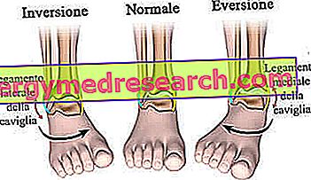
Therefore, as in the case of plantarflexion and dorsiflexion, even eversion and inversion are two opposite movements.
- Flexion and extension of the leg
The terms flexion and extension indicate two opposite movements, which vary the angle present between two adjacent anatomical segments.
In the case in question, they particularly affect the flexion and extension of the leg and the flexion and extension of the toes.
Leg. Performing a flexion movement of the leg means reducing the posterior angle, existing between the leg and the thigh.
On the other hand, performing an extension movement means increasing the posterior angle, present between the leg and the thigh.
At this point, it is important to clarify two aspects: the first is that the gesture of extending the leg is always subsequent to one of bending; the second is that, when adjusting the two gestures, the knee joint plays a decisive role. The knee allows flexion of the leg backwards and its extension until it is in line with the thigh (point of maximum extension).
Toes. Flexing the toes means bending them towards the sole of the foot (or down); vice versa, extending the toes means bending them, as far as possible, upwards.


Figure: flexion and extension of the leg. From the site: teachmeanatomy.info
FUNCTIONS OF MUSCLE FLEXORS
The gastrocnemius participates in the plantarflexion of the foot and in the flexion of the leg on the thigh.
The soleus plays a key role in the plantarflexion of the foot, especially during the execution of the race.
The gracile plantar supports gastrocnemius and soleus, in the movement of plantarflexion and flexion of the leg on the thigh.
The popliteus participates in the flexion of the leg on the thigh and in the rotation towards the inside of the tibia.
The long flexor of the big toe contributes to the plantarflexion and allows the flexion of the phalanges of the big toe (NB: during flexion, the first finger points downwards).
Finally, the long flexor of the fingers contributes to the plantarflexion and allows the flexion of the phalanges of the last 4 fingers (NB: as in the previous case, during the flexion, the last 4 fingers point downwards).
FUNCTIONS OF EXTENDED MUSCLES
The long extender of the toes has two tasks:
- It allows to extend the phalanges of the last 4 fingers, towards the back of the foot. Therefore, thanks to this muscle, the fingers tend to point upwards.
- It contributes to the dorsiflexion movement, with a slight rotation towards the outside of the foot (slight eversion).
Similarly, even the long extensor of the big toe covers two functions:
- It allows to extend the phalanges of the big toe, towards the back of the foot. As a result, the first toe tends to point upwards.
- It contributes to the dorsiflexion movement, with a slight rotation towards the inside of the foot (slight inversion).
FUNCTIONS OF ADDUCTOR MUSCLES
The tibialis anterior contributes to the dorsiflexion, while the tibialis posterior to the plantarflexion.
Both participate in the reversal movement of the foot.
FUNCTIONS OF MUSCLE ABDUCTORS
The third peroneus contributes to dorsiflexion and subversion of the foot.
The long peroneal takes part in plantarflexion and inversion. Furthermore, it accentuates the concavity of the arch (or vault) of the foot. The arch is the concave surface, located on the sole of the foot, which prevents the latter from resting completely on the ground.
Finally, the short peroneal supports the movement of plantarflexion and eversion of the foot.
Associated diseases
Like most muscles in the human body, the leg muscles can also experience contractures, strains, tears and tendon inflammation / injury.
These injuries usually affect active people, such as those who practice sports.
Among the muscular elements present in the leg, the ones that suffer most from contractures, stretching and tearing are the gastrocnemius and the soleus.
As for tendon sections, the tendon of the leg muscles most prone to injury is, of course, the Achilles tendon.