Generality
Crouzon syndrome is a rare genetic disorder that determines the presence of craniosynostosis and other rather unusual facial anomalies.
The causes of its appearance are certain alterations of the DNA that constitutes the FGFR2 and FGFR3 genes; these genetic elements are involved in the process of bone maturation during embryonic development.

The therapy consists of a series of surgical interventions, aimed at solving the most important and most dangerous symptoms.
Currently, the prognosis tends to be, very often, positive.
Recalls of genetics
Before proceeding with the description of Crouzon syndrome, it is useful to review some fundamental concepts of genetics.
What is DNA? It is the genetic heritage in which the physical features, predispositions, physical qualities, character, etc. are written. of a living organism. It is contained in all the cells of the body having a nucleus, as it is inside this one.
What are chromosomes? According to the definition, chromosomes are the structural units in which DNA is organized. The human cells contain, in their nucleus, 23 pairs of homologous chromosomes (22 of an autosomal non-sexual type and a couple of sexual type); each pair is different from another, as it contains a specific gene sequence.
What are genes? They are short stretches, or sequences, of DNA with a fundamental biological meaning: from them, in fact, proteins are derived, that is, fundamental biological molecules for life. In genes, there is "written" part of what we are and what we will become.
Each gene is present in two versions, the alleles: an allele is of maternal origin, then transmitted by the mother; the other allele is of paternal origin, therefore transmitted by the father.
What is a genetic mutation? It is an error within the DNA sequence that forms a gene. Because of this error, the resulting protein is defective or completely absent. In both cases, the effects can be deleterious both for the life of the cell, in which the mutation occurs, and for that of the organism in its totality. Congenital diseases and neoplasms (or tumors) are responsible for one or more genetic mutations.
What is Crouzon syndrome
Crouzon syndrome is a rare genetic condition characterized by craniosynostosis and an unnatural development of some facial elements, including eyes, nose, jaw and jaw.
It is a congenital disease, whose typical characteristics can already be seen during the first moments of life.
MEANING OF CRANIOSINOSTOSIS
Craniosynostosis is the term by which doctors indicate the premature fusion of one or more cranial sutures.

From the site: thecraniofacialcenter.com
Cranial sutures are the fibrous joints that bind together the bones of the cranial vault (ie the frontal, temporal, parietal and occipital bones).
In normal conditions, the fusion of cranial sutures occurs in the post-natal period (some processes even end at the age of 20). This long process of fusion allows the brain to grow and develop properly.
If, as in the case of craniosynostosis, fusion takes place too early - therefore during prenatal, perinatal * or early childhood - the encephalic elements (brain, cerebellum and brainstem) and some sense organs (eyes in particular) undergo an alteration of shape and growth.
ORIGIN OF THE NAME
Crouzon syndrome owes its name to the French physician Octave Crouzon, who has the merit of having first described the main clinical features.
Crouzon lived between the end of the 1800s and the first decades of the 1900s, exactly from 1874 to 1938. Initially, to define the syndrome that later took its name, he used the term craniofacial dysostosis.
Causes
Crouzon syndrome arises following a mutation of the FGFR2 gene, located on chromosome 10, or of the FGFR3 gene, located on chromosome 4 .
FGFR is the English acronym of Fibroblast Growth Factor Receptor, which translated into Italian is: Receptor for Fibroblast Growth Factor .
The functional role of the FGFR2 and FGFR3 genes is to produce, each, a receptor protein, with the task of regulating the maturation and embryonic development of bone tissue.
According to the researchers' theories, the mutations of FGFR2 and FGFR3 would hyper-stimulate these same genes, which, once more active, would induce an early maturation of some bone tissues, including those constituting the skull.
GENETICS
Genetic mutations responsible for Crouzon syndrome may be hereditary or may arise spontaneously after conception .
In the first case, the morbid condition - which doctors also call hereditary Crouzon syndrome - has all the characteristics of a genetic disease with an autosomal dominant transmission (or dominant inherited disease ). For an inexperienced reader of genetics, this means that:
- The disease and its symptoms are also manifested in the presence of only one mutated gene allele (it does not matter whether it comes from the mother or the father), since the latter is dominant over the healthy one.
- A parent carrying the mutation is sufficient to have the disease in part of the offspring.
- The probability that a sick child will be born, from a couple where only one of the two components carries the mutation, is 50%.
In the second case, instead, the morbid condition - which the experts indicate with the terminology of non-hereditary Crouzon syndrome - is the result of a sporadic anomalous event, which alters DNA during embryonic growth of the fetus.
Summary of the meaning of the terms hereditary, autosomal and dominant
- Hereditary: means that parents transmit the genetic alteration responsible for the disease to their offspring (ie their children).
- Autosomal: means that the mutation responsible for the disease lies in a non-sexual chromosome, therefore autosomal.
- Dominant: means that the disease causes symptoms and signs even when only one allele of the responsible gene is mutated. In simpler terms, it is as if the allele with the mutation had more power than the healthy allele.
Epidemiology
According to some estimates of the incidence rate of Crouzon syndrome, one child per approximately 60, 000 would be born with this rare condition.
Crouzon syndrome accounts for 4.5% of craniosynostosis cases.
Symptoms and Complications
Patients with Crouzon syndrome have a very specific symptomatic picture, which generally consists of:
- Problems related to craniosynostosis, including:
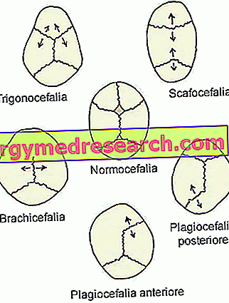
From //en.wikipedia.org/wiki/Plagiocephaly Brachicefalia, which is the crushing of the back of the head. This leads to premature fusion of the coronal cranial sutures (coronal craniosynostosis).
If left untreated, it can affect the growth of the brain and the development of cognitive abilities.
They represent an alternative to brachycephaly: trigonocephaly (fusion of the metopic suture), dolichocephaly (fusion of the sagittal suture) and plagiocephaly (fusion of the coronal sutures).
- Exophthalmos, which is the term for the protrusion of eyeballs. It could imply the presence of sight problems.
- Ocular hypertelorism, that is, eyes exaggeratedly distant from each other. With exophthalmos, it can worsen visual problems.
- Deformed nose, generally beak-shaped. If severe or untreated, this abnormality may result in respiratory problems or the same symptoms as obstructive sleep apnea syndrome.
- Increase in intracranial pressure. It is a circumstance also known as intracranial hypertension. Its presence is explained by the fact that brain structures do not have the right space to grow.
Usually known as mid-late childhood, intracranial hypertension is a potential cause of headache, vomiting and eye pain.
- Hydrocephalus, which is the result of an increase in the cerebrospinal fluid, contained in the subarachnoid space and in the cerebral ventricles.
- Arnold-Chiari malformation (or Arnold-Chiari syndrome). It is a deformity based at the base of the skull.
* Hydrocephalus and Arnold-Chiari malformation are generally two complications that arise in the absence of adequate treatments.
- Abnormalities of the mandible and maxilla .
The first is smaller than normal, while the second tends to protrude outwards. All this modifies the shape of the palate and the dental scaffolding (absence of some teeth, etc.), with repercussions (sometimes even serious) on phonation and chewing.
Some patients are born with cleft lip (cleft lip) or cleft palate.
- Hearing problems .
55% of patients with Crouzon syndrome are born without auditory canals or large abnormalities against them. This results in an absent or strongly reduced acoustic capacity.
Some subjects develop a set of hearing problems in adulthood, due to the typical clinical picture of Ménière's syndrome.
- Joint problems in the neck .
These concern 30% of Crouzon syndrome cases.
- Skin anomalies .
Patients with Crouzon syndrome supported by mutated FGFR3 have acanthosis nigricans, a dermatosis characterized by an increase in thickness (hyperkeratosis) and a darkening (hyperpigmentation) of the skin.

Two other anatomical anomalies, associated (albeit rarely) with Crouzon syndrome
- Patent ductus arteriosus
- Coarctation of the aorta
CROUZON SYNDROME AND INTELLECTIVE QUOTIENT
Thanks also to the current possibilities of curing craniosynostosis, today 97% of patients with Crouzon syndrome have normal intelligence.
Diagnosis
An experienced pediatrician may be able to diagnose Crouzon syndrome by physical examination only.
In the presence of any doubt or perplexity, it is essential to arrive at a precise conclusion:
- Radiological images, provided by X-rays or CT scan to the head
- A genetic test, aimed at finding possible DNA mutations.
EXAMINATION OBJECTIVE
The objective examination consists in the accurate analysis of the head and the anomalies present on this one.
Cranial deformities induced by craniosynostosis (for example brachycephaly) are among the most characteristic clinical signs of Crouzon syndrome and on which the physician bases part of his diagnostic conclusions.
RADIOLOGICAL TESTS
X-rays and CT at the head show which cranial sutures fused prematurely.
The craniosynostosis that characterizes Crouzon syndrome involves coronal sutures, so a finding of fusion at the level of the latter is very often crucial information for diagnostic purposes.
GENETIC EXAMINATION
In addition to showing whether the DNA has mutations, the genetic test identifies the exact gene that causes Crouzon syndrome, whether FGFR2 or FGFR3.
Treatment
Today, people with Crouzon syndrome can rely on various treatments, depending on the severity of the condition and the symptoms.
In fact, the doctors have taken steps to guarantee:
- Surgery for the resolution of craniosynostosis and its symptoms.
- Acoustic supports, in case of hearing problems.
- Therapies for improving language skills.
- Surgical therapies to improve jaw and mandible abnormalities.
- A surgery, known as a tracheostomy, for the resolution of respiratory problems.
Please note: Crouzon syndrome is a morbid condition that results from a genetic alteration of DNA that cannot be cured. Therefore, in fact, doctors treat the disease only from a symptomatic point of view.
SURGERY FOR CRANIOSINOSTOSIS
There are two therapeutic goals for surgery:
- Provide the encephalic structures and the eyes with that space, which serves them to develop and function at its best.
- Give the head a normal shape, then solve the problem of brachycephaly.
Surgeons have the opportunity to perform the surgery in two different ways (or approaches): through a traditional surgery operation - also called "open-air" - or through an endoscopic surgery operation.
The "open-air" operation involves making an incision on the head, through which the operating physician extracts the malformed bone or cranial bones and which must be remodeled. At the end of the remodeling, the surgeon reinserts the previously extracted bone structures and closes the incision with stitches.
The endoscopic surgery intervention, instead, involves the use of an endoscope and the practice of a very small incision on the head, through which the operating doctor inserts the endoscope itself.
The endoscope is actually a thin and flexible tube, equipped with an optical fiber camera (in the end inserted into the skull) and connected to a monitor. Through this particular instrument and the images that it projects on the monitor, the surgeon is able to separate the fused cranial sutures prematurely, with remarkable precision and without resorting to skin incisions and bone extraction.
According to the experts, the best time to perform the operation is during early childhood (first 12 months of life), as the bones are more easily shaped.
However, it should be remembered that the lower the age of the patient, the higher the risk of a recasting of the same cranial sutures (recurrence). In case of recurrence, the surgical operation must be repeated.
According to some statistical researches, 10-20% of very young subjects, subjected to craniosynostosis surgery, need to undergo a second operation, due to a relapse.
TREATMENT OF ACOUSTIC PROBLEMS
In addition to prescribing the use of acoustic supports, doctors also recommend periodic audits of the hearing faculties, as this is the best way to prevent any worsening of the problems in progress.
SURGICAL THERAPIES FOR JAWS AND MANDIBLE ANOMALIES
The treatment of maxillary and mandibular anomalies includes surgery for the realignment of the maxilla and / or jaw, some dental care for the arrangement of the dental arches and the operation for the resolution of the cleft lip and / or cleft palate.
TRACHEOSTOMY
The tracheostomy is the surgical operation by which the doctor creates, at the level of the neck (where the trachea passes), a passageway for the air destined for the lungs. This allows those who undergo this operation to breathe again correctly.
To convey the air into the lungs, you need a small tube, called a trascheostomy tube, which is the right size for insertion into the trachea.
Prognosis
In general, the prognosis depends on the severity of the craniosynostosis: if the latter is curable with good results, patients with Crouzon syndrome can enjoy an almost normal life.