Generality
The ulna is the even bone which, together with the radius (with respect to which it is in the medial position), composes the skeleton of each forearm.

To simplify the study, the anatomy experts divide it into three portions: the proximal end (or proximal epiphysis), the body (or diaphysis) and the distal end (or distal epiphysis).
The proximal end is the bone portion closest to the humerus and with which it articulates.
The body is the central portion, including the proximal epiphysis and the distal epiphysis.
Finally, the distal end is the portion adjacent to the carpal bones, which represent the first part of the skeleton of the hand.
What is Ulna
The ulna is the even bone which, together with the radium, makes up the skeleton of each forearm .
The forearm is the anatomical region of the upper limb between the upper arm and the hand below.
Belonging to the category of long bones, the ulna is the protagonist of two important joints of the human body: one with the arm bone (the humerus), called the elbow joint, and another with the carpal bones of the hand, called wrist joint .
POSITION COMPARED TO RADIO
The ulna runs parallel to the radium, in a medial position if the hand is turned with the palm towards the observer.
An explanation of the concept of media (and its opposite meaning, ie lateral) is present in the box below.
Important note: meaning of medial and lateral
Medial and lateral are two terms with the opposite meaning. However, to fully understand what they mean, it is necessary to take a step back and review the concept of the sagittal plan.
Figure: the plans with which the anatomists dissect the human body. In the image, in particular, the sagittal plane is highlighted.
The sagittal plane, or median plane of symmetry, is the antero-posterior division of the body, a division from which two equal and symmetrical halves are derived: the right half and the left half. For example, from a sagittal plane of the head derive a half, which includes the right eye, the right ear, the right nasal nostril and so on, and a half, which includes the left eye, the left ear, the left nasal nostril etc.
Returning to the medial-lateral concepts, the word media indicates a relationship of proximity to the sagittal plane; while the word side indicates a relationship of distance from the sagittal plane.
All anatomical organs can be medial or lateral with respect to a reference point. A couple of examples clarify this statement:
First example. If the reference point is the eye, it is lateral to the nasal nostril of the same side, but medial to the ear.
Second example. If the reference point is the second toe, this element is lateral to the first toe (toe), but medial to all the others.
IN THE LOWER ARTS IT CORRESPONDS TO ...
In the lower limb, the bone corresponding to the ulna is the fibula . Together with the tibia, the fibula constitutes the skeleton of the leg . Like the ulna, fibula and tibia are two even bones.
Anatomy
The anatomists identify three main bone regions (or portions) in the ulna: the proximal end (or proximal epiphysis), the body (or diaphysis) and the distal end (or distal epiphysis).
Anatomical meaning of proximal and distal
Proximal and distal are two terms with opposite meaning.Proximal means "closer to the center of the body" or "closer to the point of origin". Referring to the femur, for example, it indicates the portion of this bone closest to the trunk.
Distal, on the other hand, means "farther from the center of the body" or "farther from the point of origin". Referred (always to the femur), for example, it indicates the portion of this bone furthest from the trunk (and closer to the knee joint).
End? PROXIMAL OF ULNA
The proximal end of the ulna is the bone portion closest to the arm and which, joining the latter's bone (the humerus ), forms the elbow joint.
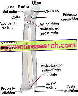
The terminal and initial site of several muscles of the upper limb, the proximal epiphysis of the ulna has some particularly relevant anatomical structures, including: the olecranon, the coronoid process, the semilunar groove, the trochlear recess, the hollow radial and tuberosity of the ulna.
- Olecrano . Most proximal part of the ulna, it is a hook-shaped bone projection that serves for the constitution of the elbow joint. In fact it contributes to the formation of the trochlear recess, which - as will be seen later - houses the trochlea of the humerus.
As a hook, the olecranon is concave anteriorly and convex posteriorly.
It houses the terminal heads of some muscles (for example the anconeus muscle) and the initial heads of others (for example the flexor carpi ulnar muscle).
- Coronoid process . It is a sort of bone crest, located on the anterior surface of the ulna and projected forward. It contributes to the formation of the trochlear recess, therefore it serves to complete the elbow joint.
The coronoid process receives the initial heads of different muscles of the hand (flexor muscles of the fingers of the hand, both superficial and deep).
- Trochlear recess (or semi-lunar notch ). It is a depression, which, when viewed from the side, together with the olecranon and the coronoid process, looks like a wrench. As anticipated, the function of the trochlear cavity is to articulate with the humeral trochlea and generate the articulation of the elbow. Equipped with a smooth surface, the humeral trochlea occupies the inferior margin of the humerus, therefore it resides in the distal end of this bone.
- Radial recess . Located laterally to the trochlear recess, it is a second depression (smaller than the trochlear recess), which serves to articulate the ulna with the radium. The area of the radio, which enters into communication with the radial hollow of the ulna, is called the head of the radium.
- Tuberosity of the ulna . It is a bony prominence, immediately inferior to the coronoid process. It houses the terminal head of the brachial muscle.

BODY OF THE ULNA
The body is the central portion of the ulna, between the proximal end and the distal end.
Along its entire path, it has 3 surfaces - the anterior, posterior and medial - and 3 edges - the posterior, the interosseous and the anterior.
Among the aforementioned elements, they deserve a report:
- The anterior and posterior surfaces, as they host the initial head of several important muscles of the forearm and hand.
- The back border, since it is palpable from the beginning to the end of its path, along the entire body of the ulna.
- The interosseous border, as it engages the so-called interosseous radio-ulnar membrane . The radio-ulnar interosseous membrane is a thin sheet of fibrous tissue, interposed between radius (medially) and ulna (laterally), which indirectly unites the two bones of the forearm.
Bone unions involving interosseous membranes are a particular type of fibrous joints, identified with the name of syndesmosis .
Another important fibrous articulation, very similar to that present between radium and ulna, is the syndesmosis located between tibia and fibula.
End? DISTAL OF THE ULNA
The distal end of the ulna is the bone portion closest to the wrist and with which it contributes to the formation of the homonymous articulation.
The anatomical element that distinguishes it is the so-called ulna head .
Of rounded shape, the head of the ulna has the important task of articulating with the ulnar groove of the radium, thus uniting ulna and radium.
Furthermore, on the inferior margin, in medial position, it presents a bone projection, which acts as an insertion zone for one of the two ends of the collateral ulnar carpal ligament (NB: the carpus is the group of bones of the hand that forms the wrist) .
This bone projection is called the styloid process (of the ulna).

BLOOD SPRAYING
Internally, long bones, such as the ulna (but also the radius or humerus), have a very specific network of arteries and veins, which serves to guarantee them the right supply of oxygen and nutrients.
Arteries - that is, in vessels that carry oxygen-rich blood - are the so-called nutritive artery and periosteum arteries ; the veins - that is, the vessels that drain the oxygen-poor blood - are the so-called nutritive vein and the periosteum veins .
In the case of the ulna, the aforementioned arteries derive from the anterior interosseous artery, while the aforesaid veins flow into the anterior interosseous vein .
The nutritive artery and the nutritive vein deserve a particular note, as they penetrate the body of the ulna, through a characteristic structure of long bones: the nutritious hole .
The nutritious hole, also called the nutritious canal, is generally located in the ulna's body, roughly in the middle position.

Figure: The nutritious vessels and the nourishing hole in the long bones.
ULNA ARTICULATION
In summary, the joints in which the ulna participates are four:
- The elbow joint, fruit of the union between trochlear hollow of the ulna and trochlea of the humerus.
- The upper radio-ulnar joint, which joins the ulnar groove, located on the proximal epiphysis of the ulna, to the head of the radius, located on the proximal end of the radius.
- The inferior radio-ulnar articulation, which joins the distal end of the ulna (in this case, the head of the ulna) to the distal end of the radium (to be precise, the ulnar groove of the radium).
- The wrist joint. The ulna inserts into the ulnar collateral ligament of the carpus, which serves to weld the wrist joint.
OBSIFICATION OF THE ULNA
Three ossification centers contribute to the formation of the ulna: one centered on the body, one on the proximal epiphysis and one on the distal epiphysis.
To begin the process of ossification is the center present on the body of the ulna; to follow and in succession, the center of the distal end and the center of the proximal end enter into action.
Going into more detail:
- The ossification center of the body is activated around the 8th week of fetal life. Its activity causes the bone to develop towards the body and the ends.
- The ossification center of the distal end (in the middle of the ulna head) is set in motion at the age of about 4 years. The bone portion it gives rise to meets the bony portion of the ossification center of the body at about the 12th year of life.
- The ossification center of the proximal end (near the olecranon) begins its activity at about the 10th year of life. The resulting bone portion meets the bony portion of the body around the sixteenth year of life.
Functions
The ulna covers at least two important functions.
The first concerns his involvement in the elbow and wrist joints. These articular elements are fundamental for numerous movements and gestures of the upper limb, such as the throwing of an object, writing, lifting a weight, etc.
The second important function of the ulna is to receive the muscles of the forearm and of the hand. These muscular structures support the action of the aforementioned joints; in fact, without them, the aforementioned movements and gestures would be impossible.
| List of the main muscles that originate and end at the ulna. | ||
Muscle | Head end or initial leader | Contact site on the tibia |
| Brachial triceps muscle | Head end | Upper part of the posterior surface of the olecranon |
| Anconeus muscle | Head end | olecranon |
| Brachialis muscle | Head end | Anterior surface of the coronoid process of the ulna |
| Pronator round muscle | Initial leader | Medial surface of the coronoid process |
| Flexor carpal ulnar muscle | Initial leader | olecranon |
| Superficial flexor muscle of the fingers | Initial leader | Coronoid process |
| Deep flexor muscle of the fingers | Initial leader | Coronoid process |
| Pronator square muscle | Initial leader | Distal section of the anterior surface of the ulna body |
| Extensor ulnar carpus muscle | Initial leader | Posterior border of the ulna |
| Supinator muscle | Initial leader | Proximal portion of the ulna |
| Long abductor muscle of the thumb | Initial leader | Back surface of the ulna body |
| Long extensor muscle of the thumb | Initial leader | Back surface of the ulna body |
| Extensor index muscle | Initial leader | Distal posterior surface of the ulna body |
Diseases of the Ulna
Like practically any other bone in the human body, the ulna can fracture as a result of traumas against it.
The body of the ulna is the most subject bone portion to fracture. However, the aforesaid bone can break, with discreet frequency, also at the level of the olecranon (proximal end) and at distal level.
Moreover, the so-called Monteggia fracture and the so-called Galeazzi fracture are also relevant (NB: the proper names belong to the doctors who described, for the first time, this type of accident)
MONTEGGIA FRACTURE AND GALEAZZI FRACTURE
Monteggia 's fracture and Galeazzi's fracture are two injuries that have a particular characteristic in common: they affect both the bones of the forearm, so both ulna and radius.
To cause the double rupture of ulna and radium, they are traumas whose impact force is transmitted from bone to bone, through the interosseous membrane.
Monteggia fracture is a clinical condition characterized by rupture of the ulna's body and dislocation of the radial head (NB: unlike the ulna, the radial head is a bone region located on the proximal epiphysis).
Galeazzi's fracture, on the other hand, is a fracture of the distal end of the radius, combined with a dislocation of the ulna's head .