Category exams

Sacral Loin Magnetic Resonance
exams

Sacral Loin Magnetic Resonance

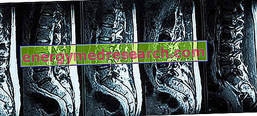
Generality The sacral loin magnetic resonance is the important diagnostic tool that allows to visualize, in a detailed way, the terminal extremity of the vertebral column (from the lumbar tract to the coccyx). Through the sacral loin magnetic resonance, the radiologist is able to: formulate diagnoses of pathologies, such as for example the herniated disc, sciatica, spinal tumors or discopathies; tracing back to the causes of persistent pain in the lumbar area of ​​the back; to investigate the severity of congenital malformations of the spine, such as spina bifida; etc. Un

Read More
exams

Barium sulfate

What is that Barium sulfate is a substance used as a contrast agent in the performance of certain types of diagnostic tests, such as CT scans and radiographs. From the chemical point of view, barium sulfate is an inorganic salt (chemical formula BaSO 4 ), which appears as a white powder that is scarcely soluble both in water and in organic solvents
Read More
exams

Diet for Colonoscopy

Importance of Preparation The diet is an integral part of the preparation for colonoscopy, regardless of whether it is the traditional optical exam or the virtual one. A good preparation for colonoscopy is made up of three key elements: possible adjustment of pharmacological therapies in place and adoption of other prophylactic measures; low fiber diet ; use of drugs with laxative effect
Read More
exams

What is the fundus exam?

The examination of the fundus fund is one of the routine diagnostic tests carried out by the eye doctor. It is an analysis of the fundus of the eye (the posterior part of the eyeball) that allows to detect the presence of pathologies, such as retinal detachment or age-related macular degeneration . How to do it The examination of the fundus is carried out by using some equipment that does not involve invasive procedures for the patient
Read More
exams

Risks of the RX-chest: is it an invasive exam?

During the diagnostic procedure called RX-thorax or chest radiography, the production of the thoracic image takes place by means of an instrument that emits high-energy ionizing radiations. Are these radiations dangerous? At high doses, ionizing radiations are harmful to all living beings, while, at very low doses as in the case of an X-ray chest, they are dangerous only if the exposure is very prolonged or repeated regularly throughout life
Read More
exams

Under what circumstances is an RX-chest performed?

The chest X -ray , also known as chest radiograph , is a widely practiced diagnostic test, which has the purpose of reproducing the organs and bone structures of an individual's chest on a photographic plate (or a digital detector). Generally, it is performed when an individual suffers from dyspnea and possibly even severe and / or persistent cough, chest pain, chest pain due to trauma, fever
Read More
exams

What is chest radiography and how is it performed?

The chest X -ray , also known as chest radiograph , is a diagnostic test that produces images of the heart, lungs, respiratory airways, sternum bones, back bones and blood vessels, present in the thoracic area. The production of images is possible thanks to a particular technological tool, which emits ionizing radiation
Read More
exams

What is tonometry?

Tonometry consists in measuring the internal pressure of the eye - which under normal conditions must be between 10 and 20 millimeters of mercury (mmHg) - and allows the risk of glaucoma to be verified. How to do it For this exam it is possible to use basically two types of instruments, the contact tonometer and the blow tonometer
Read More
exams

Optical coherence tomography

Optical coherence tomography (OCT - acronym of "Optical Coherent Tomography" ) is a very innovative imaging technique , which "takes" high-resolution photographs of the retinal tissue , providing information on the thickness, conformation and relationship between various layers that make it up
Read More
exams

How many radiations does a RX-chest or a thoracic CT emit?

Radiological diagnostic tests, such as RX-thorax or thoracic CT , produce internal body images, thanks to equipment that emits different doses of ionizing radiation. But how is radioactivity measured and what is the precise amount of ionizing radiation that affects the patient during these tests? First of all, the most commonly used unit of measurement for the quantification of radioactivity is the millisievert ( mSv )
Read More
exams

Magnetic resonance imaging

Generality Magnetic resonance imaging is a diagnostic test that allows the visualization of the inside of our body without performing surgical operations or administering dangerous ionizing radiation. Conceived and developed around 1980, magnetic resonance has undergone a constant process of technological evolution over the years
Read More